Onishi Masazumi, Takahashi Kazuhiro, Maeda Fumihiko, Akasaka Toshihide
Department of Dermatology, Iwate Medical University, Morioka, Japan.
Case Rep Dermatol. 2015 Dec 17;7(3):352-7. doi: 10.1159/000442704. eCollection 2015 Sep-Dec.
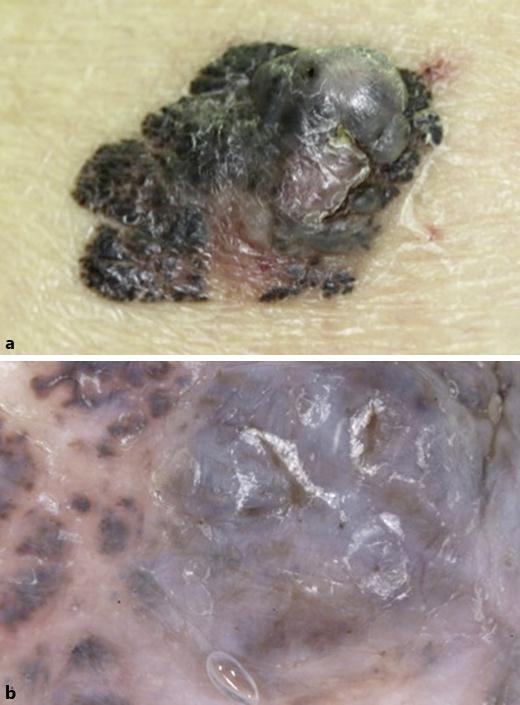
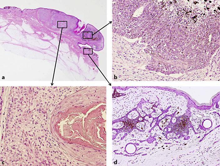
A 70-year-old Japanese man presented at our hospital with an asymptomatic, blackish, irregularly shaped plaque with a gray nodule in the periphery on his left lower leg. The lesion had been present for 10 years and had recently enlarged, associated with bleeding. Histopathologically, the tumor consisted of three distinct parts: The first part showed massive aggregation of basophilic basaloid cells with peripheral palisading and abundant melanin granules, and was diagnosed as solid-type basal cell carcinoma. The second part showed aggregation of clear cells with squamous eddies, and was diagnosed as proliferating trichilemmal tumor. The third part showed reticular aggregation of basaloid cells with infundibular cysts in the papillary dermis, and was diagnosed as infundibulocystic basal cell carcinoma. We diagnosed this tumor as basal cell carcinoma with various forms of hair follicle differentiation, including differentiation into the outer root sheath.
一名70岁的日本男性因左小腿出现无症状、黑色、形状不规则的斑块,周边有灰色结节而到我院就诊。该病变已存在10年,近期增大并伴有出血。组织病理学检查显示,肿瘤由三个不同部分组成:第一部分表现为嗜碱性基底样细胞大量聚集,周边呈栅栏状排列,并有丰富的黑色素颗粒,诊断为实性基底细胞癌;第二部分表现为透明细胞聚集并伴有鳞状漩涡,诊断为增生性毛鞘瘤;第三部分表现为基底样细胞呈网状聚集,乳头真皮层有漏斗状囊肿,诊断为漏斗状囊型基底细胞癌。我们将该肿瘤诊断为具有多种毛囊分化形式的基底细胞癌,包括向毛外根鞘分化。