• 文献检索
  • 文档翻译
  • 深度研究
  • 学术资讯
  • Suppr Zotero 插件Zotero 插件
  • 邀请有礼
  • 套餐&价格
  • 历史记录
应用&插件
Suppr Zotero 插件Zotero 插件浏览器插件Mac 客户端Windows 客户端微信小程序
定价
高级版会员购买积分包购买API积分包
服务
文献检索文档翻译深度研究API 文档MCP 服务
关于我们
关于 Suppr公司介绍联系我们用户协议隐私条款
关注我们

Suppr 超能文献

核心技术专利:CN118964589B侵权必究
粤ICP备2023148730 号-1Suppr @ 2026

文献检索

告别复杂PubMed语法,用中文像聊天一样搜索,搜遍4000万医学文献。AI智能推荐,让科研检索更轻松。

立即免费搜索

文件翻译

保留排版,准确专业,支持PDF/Word/PPT等文件格式,支持 12+语言互译。

免费翻译文档

深度研究

AI帮你快速写综述,25分钟生成高质量综述,智能提取关键信息,辅助科研写作。

立即免费体验

健康眼中视乳头周围视网膜灌注与近视有关吗?一项前瞻性对照研究。

Is the peripapillary retinal perfusion related to myopia in healthy eyes? A prospective comparative study.

作者信息

Wang Xiaolei, Kong Xiangmei, Jiang Chunhui, Li Mengwei, Yu Jian, Sun Xinghuai

机构信息

Department of Ophthalmology and Visual Science, Eye, Ear, Nose and Throat Hospital, Shanghai Medical College of Fudan University, Shanghai, China.

Department of Ophthalmology and Visual Science, Eye, Ear, Nose and Throat Hospital, Shanghai Medical College of Fudan University, Shanghai, China Key Laboratory of Myopia, Ministry of Health (Fudan University), Shanghai, China Shanghai Key Laboratory of Visual Impairment and Restoration (Fudan University), China.

出版信息

BMJ Open. 2016 Mar 11;6(3):e010791. doi: 10.1136/bmjopen-2015-010791.

DOI:10.1136/bmjopen-2015-010791
PMID:26969645
原文链接:https://pmc.ncbi.nlm.nih.gov/articles/PMC4800142/
Abstract

OBJECTIVES

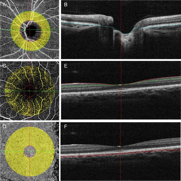
The aim of this study was to evaluate the peripapillary and parafoveal perfusion of young, healthy myopic subjects with spectral domain optical coherence tomography (OCT) angiography.

DESIGN

A prospective comparative study was conducted from December 2014 to January 2015.

SETTING

Participants recruited from a population-based study performed by the Eye, Ear, Nose and Throat Hospital of Fudan University in Shanghai.

PARTICIPANTS

A total of 78 Chinese normal subjects (78 eyes) with different refraction were included. Myopia was divided into 4 groups on the basis of the refractive status: 20 eyes with emmetropia (mean spherical equivalent (MSE) 0.50D to -0.50D), 20 eyes with mild myopia (MSE -0.75D to -2.75D), 20 eyes with moderate myopia (MSE -3.00D to -5.75D), and 18 eyes with high myopia (MSE≤-6.00D).

MAIN OUTCOME MEASURES

Peripapillary and parafoveal retinal and choroidal perfusion parameters and their relationships with axial length (AL) and retinal nerve fibre layer (RNFL) thickness were analysed.

RESULTS

Significant differences were found for the retinal flow index and vessel density in the peripapillary area among the 4 groups, but not in the parafoveal area. The high myopia group had the lowest peripapillary retinal flow index and vessel density. In addition, there was a negative correlation (β=-0.002, p=0.047) between the AL and peripapillary retinal flow index and a positive correlation between RNFL thickness and the peripapillary retinal perfusion parameters (flow index: β=0.001, p=0.006; vessel density: β=0.350, p=0.002) even after adjustment for other variables.

CONCLUSIONS

Highly myopic eyes have a decreased peripapillary retinal perfusion compared with emmetropic eyes. Such vascular features might increase the susceptibility to vascular-related eye diseases.

摘要

目的

本研究旨在利用频域光学相干断层扫描(OCT)血管造影术评估年轻健康近视受试者的视乳头周围和黄斑旁灌注情况。

设计

2014年12月至2015年1月进行了一项前瞻性对照研究。

地点

参与者来自复旦大学附属眼耳鼻喉科医院开展的一项基于人群的研究。

参与者

共纳入78名具有不同屈光状态的中国正常受试者(78只眼)。根据屈光状态将近视分为4组:20只正视眼(平均球镜当量(MSE)0.50D至 -0.50D),20只轻度近视眼(MSE -0.75D至 -2.75D),20只中度近视眼(MSE -3.00D至 -5.75D),以及18只高度近视眼(MSE≤-6.00D)。

主要观察指标

分析视乳头周围和黄斑旁视网膜及脉络膜灌注参数及其与眼轴长度(AL)和视网膜神经纤维层(RNFL)厚度的关系。

结果

4组之间视乳头周围区域的视网膜血流指数和血管密度存在显著差异,但黄斑旁区域无差异。高度近视组的视乳头周围视网膜血流指数和血管密度最低。此外,即使在对其他变量进行校正后,AL与视乳头周围视网膜血流指数之间仍存在负相关(β=-0.002,p=0.047),RNFL厚度与视乳头周围视网膜灌注参数之间存在正相关(血流指数:β=0.001,p=0.006;血管密度:β=0.350,p=0.002)。

结论

与正视眼相比,高度近视眼的视乳头周围视网膜灌注减少。这种血管特征可能会增加患血管相关性眼病的易感性。

https://cdn.ncbi.nlm.nih.gov/pmc/blobs/0174/4800142/5c2b1aa86e08/bmjopen2015010791f01.jpg

相似文献

1
Is the peripapillary retinal perfusion related to myopia in healthy eyes? A prospective comparative study.健康眼中视乳头周围视网膜灌注与近视有关吗?一项前瞻性对照研究。
BMJ Open. 2016 Mar 11;6(3):e010791. doi: 10.1136/bmjopen-2015-010791.
2
Vascular flow density in pathological myopia: an optical coherence tomography angiography study.病理性近视中的血管血流密度:一项光学相干断层扫描血管造影研究。
BMJ Open. 2017 Feb 3;7(2):e013571. doi: 10.1136/bmjopen-2016-013571.
3
Assessment of superficial retinal microvascular density in healthy myopia.健康近视患者视网膜浅层微血管密度的评估
Int Ophthalmol. 2019 Aug;39(8):1861-1870. doi: 10.1007/s10792-018-1014-z. Epub 2018 Sep 3.
4
Effect of refractive status on peripapillary retinal nerve fibre layer thickness: a study by RTVue spectral domain optical coherence tomography.屈光状态对视盘周围视网膜神经纤维层厚度的影响:应用 RTVue 谱域光学相干断层扫描的研究。
Br J Ophthalmol. 2013 Jan;97(1):75-9. doi: 10.1136/bjophthalmol-2012-301865. Epub 2012 Nov 10.
5
The Characteristics of Peripapillary Retinal Perfusion by Optical Coherence Tomography Angiography in Tessellated Fundus Eyes.视网膜呈镶嵌状眼底眼中光学相干断层扫描血管造影显示的视乳头周围视网膜灌注特征
PLoS One. 2016 Jul 27;11(7):e0159911. doi: 10.1371/journal.pone.0159911. eCollection 2016.
6
[Analysis of the correlation between peripapillary retinal nerve fiber layer cross-sectional area and myopia in individuals aged 50 years and above].[50岁及以上人群视乳头周围视网膜神经纤维层横截面积与近视的相关性分析]
Zhonghua Yan Ke Za Zhi. 2023 Jul 11;59(7):550-556. doi: 10.3760/cma.j.cn112142-20221227-00660.
7
Macular and peripapillary vessel density in myopic eyes of young Chinese adults.中国年轻近视人群的黄斑区及视盘周围血管密度。
Clin Exp Optom. 2020 Nov;103(6):830-837. doi: 10.1111/cxo.13047. Epub 2020 Feb 12.
8
Optical coherence tomography angiography of superficial retinal vessel density and foveal avascular zone in myopic children.近视儿童的浅层视网膜血管密度和中心无血管区的光相干断层扫描血管造影。
PLoS One. 2019 Jul 18;14(7):e0219785. doi: 10.1371/journal.pone.0219785. eCollection 2019.
9
The Relationship between Macular Vessel Density and Thickness with Light Sensitivity in Myopic Eyes.近视眼中黄斑血管密度和厚度与光敏感度的关系。
Curr Eye Res. 2019 Oct;44(10):1104-1111. doi: 10.1080/02713683.2019.1627461. Epub 2019 Jun 13.
10
Influence of axial length on peripapillary retinal nerve fiber layer thickness in children: a study by RTVue spectral-domain optical coherence tomography.眼轴长度对儿童视乳头周围视网膜神经纤维层厚度的影响:应用 RTVue 谱域光学相干断层扫描的研究。
Curr Eye Res. 2013 Dec;38(12):1241-7. doi: 10.3109/02713683.2013.820328. Epub 2013 Aug 23.

引用本文的文献

1
Retinal and Choroidal Vascular Biomarkers Are Correlated With the Degree of Myopia.视网膜和脉络膜血管生物标志物与近视程度相关。
Transl Vis Sci Technol. 2025 Aug 1;14(8):39. doi: 10.1167/tvst.14.8.39.
2
Research on the correlation between retinal vascular parameters and axial length in children using an AI-based fundus image analysis system.使用基于人工智能的眼底图像分析系统研究儿童视网膜血管参数与眼轴长度之间的相关性。
PLoS One. 2025 Jun 17;20(6):e0324352. doi: 10.1371/journal.pone.0324352. eCollection 2025.
3
Repeatability of Fractal Analysis in OCT Angiography of the Macula and Optic Nerve Head.

本文引用的文献

1
Correlation between optic disc perfusion and glaucomatous severity in patients with open-angle glaucoma: an optical coherence tomography angiography study.开角型青光眼患者视盘灌注与青光眼严重程度的相关性:一项光学相干断层扫描血管造影研究。
Graefes Arch Clin Exp Ophthalmol. 2015 Sep;253(9):1557-64. doi: 10.1007/s00417-015-3095-y. Epub 2015 Aug 11.
2
Macular perfusion in healthy Chinese: an optical coherence tomography angiogram study.健康中国人的黄斑灌注:一项光学相干断层扫描血管造影研究。
Invest Ophthalmol Vis Sci. 2015 May;56(5):3212-7. doi: 10.1167/iovs.14-16270.
3
Optical coherence tomography angiography of optic disc perfusion in glaucoma.
黄斑和视神经乳头光学相干断层扫描血管造影中分数维分析的可重复性
Ophthalmol Sci. 2025 Apr 3;5(5):100784. doi: 10.1016/j.xops.2025.100784. eCollection 2025 Sep-Oct.
4
Sectoral Changes in Neuroretinal Rim Pallor Across Refractive Error.不同屈光不正患者神经视网膜边缘苍白的扇形变化
Ophthalmol Sci. 2025 Jan 7;5(3):100705. doi: 10.1016/j.xops.2025.100705. eCollection 2025 May-Jun.
5
Comparative analysis of the optic nerve microvasculature between different optic disc phenotypes of normal-tension glaucoma patients.正常眼压性青光眼患者不同视盘表型之间视神经微血管的比较分析。
BMC Ophthalmol. 2025 Mar 26;25(1):152. doi: 10.1186/s12886-025-03987-z.
6
Association of myopia and parapapillary choroidal microvascular density in primary open-angle glaucoma.原发性开角型青光眼患者近视与视乳头旁脉络膜微血管密度的相关性
PLoS One. 2025 Feb 25;20(2):e0317881. doi: 10.1371/journal.pone.0317881. eCollection 2025.
7
Retinal capillary density among healthy Egyptian and South Asian students: an optical coherence tomography angiography study.健康埃及和南亚学生的视网膜毛细血管密度:一项光学相干断层扫描血管造影研究。
Int J Ophthalmol. 2025 Jan 18;18(1):111-116. doi: 10.18240/ijo.2025.01.13. eCollection 2025.
8
Retinal nerve fiber layer thickness and radial peripapillary capillaries density in myopic adults with optical coherence tomography angiography.近视成年人的光学相干断层扫描血管造影的视网膜神经纤维层厚度和视盘周围毛细血管密度。
BMC Ophthalmol. 2024 Sep 18;24(1):407. doi: 10.1186/s12886-024-03673-6.
9
Peripheral Superficial Retina Vascular Density and Area of Radial Peripapillary Capillaries Changes in Myopic Individuals: A Wide-Field OCT Angiography Study.近视患者周边浅层视网膜血管密度和放射状视神经周围毛细血管面积的变化:广角 OCT 血管造影研究。
Transl Vis Sci Technol. 2024 Sep 3;13(9):21. doi: 10.1167/tvst.13.9.21.
10
Effect of magnification error and axial length on circumpapillary capillary density and retinal nerve fiber layer thickness.放大误差和眼轴长度对周边视网膜毛细血管密度和神经纤维层厚度的影响。
Sci Rep. 2024 Aug 14;14(1):18874. doi: 10.1038/s41598-024-69864-9.
青光眼视盘血流的光学相干断层扫描血管造影。
Ophthalmology. 2014 Jul;121(7):1322-32. doi: 10.1016/j.ophtha.2014.01.021. Epub 2014 Mar 12.
4
Parafoveal retinal vascular response to pattern visual stimulation assessed with OCT angiography.用 OCT 血管造影评估图形视觉刺激的旁中心视网膜血管反应。
PLoS One. 2013 Dec 2;8(12):e81343. doi: 10.1371/journal.pone.0081343. eCollection 2013.
5
Quantitative OCT angiography of optic nerve head blood flow.视神经乳头血流的定量光学相干断层扫描血管造影
Biomed Opt Express. 2012 Dec 1;3(12):3127-37. doi: 10.1364/BOE.3.003127. Epub 2012 Nov 7.
6
Cellular and physiological mechanisms underlying blood flow regulation in the retina and choroid in health and disease.健康和疾病状态下视网膜和脉络膜血流调节的细胞和生理机制。
Prog Retin Eye Res. 2012 Sep;31(5):377-406. doi: 10.1016/j.preteyeres.2012.04.004. Epub 2012 May 3.
7
Split-spectrum amplitude-decorrelation angiography with optical coherence tomography.光学相干断层扫描的分裂谱幅度去相关血管造影术。
Opt Express. 2012 Feb 13;20(4):4710-25. doi: 10.1364/OE.20.004710.
8
Prevalence and risk factors for refractive errors in Indians: the Singapore Indian Eye Study (SINDI).印度人屈光不正的患病率和危险因素:新加坡印度眼研究 (SINDI)。
Invest Ophthalmol Vis Sci. 2011 May 16;52(6):3166-73. doi: 10.1167/iovs.10-6210.
9
Ocular blood flow measurements in healthy human myopic eyes.健康人近视眼的眼血流测量。
Graefes Arch Clin Exp Ophthalmol. 2010 Nov;248(11):1587-94. doi: 10.1007/s00417-010-1407-9. Epub 2010 May 26.
10
Prevalence and progression of myopic retinopathy in Chinese adults: the Beijing Eye Study.中国成年人近视性视网膜病变的患病率和进展:北京眼病研究。
Ophthalmology. 2010 Sep;117(9):1763-8. doi: 10.1016/j.ophtha.2010.01.020. Epub 2010 May 5.