Calvo-Maroto Ana M, Esteve-Taboada José J, Pérez-Cambrodí Rafael J, Madrid-Costa David, Cerviño Alejandro
Optometry Research Group, Department of Optics & Optometry & Vision Sciences, University of Valencia, Burjassot, 46100 Valencia, Spain.
Department of Ophthalmology, Oftalmar, Medimar International Hospital, 03016 Alicante, Spain.
J Ophthalmol. 2016;2016:1287847. doi: 10.1155/2016/1287847. Epub 2016 Feb 10.
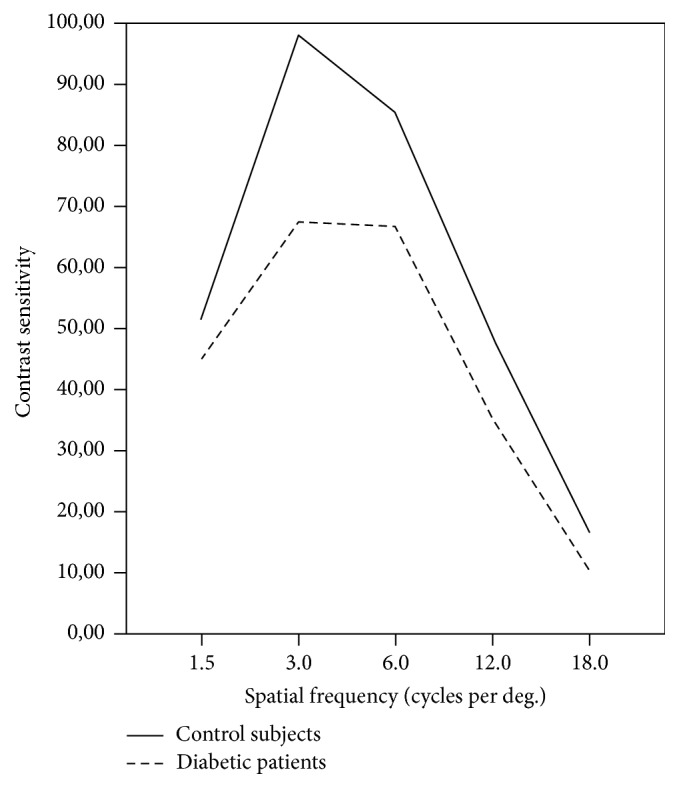
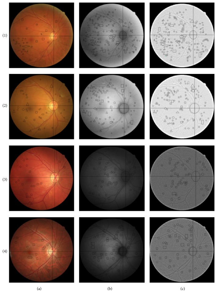
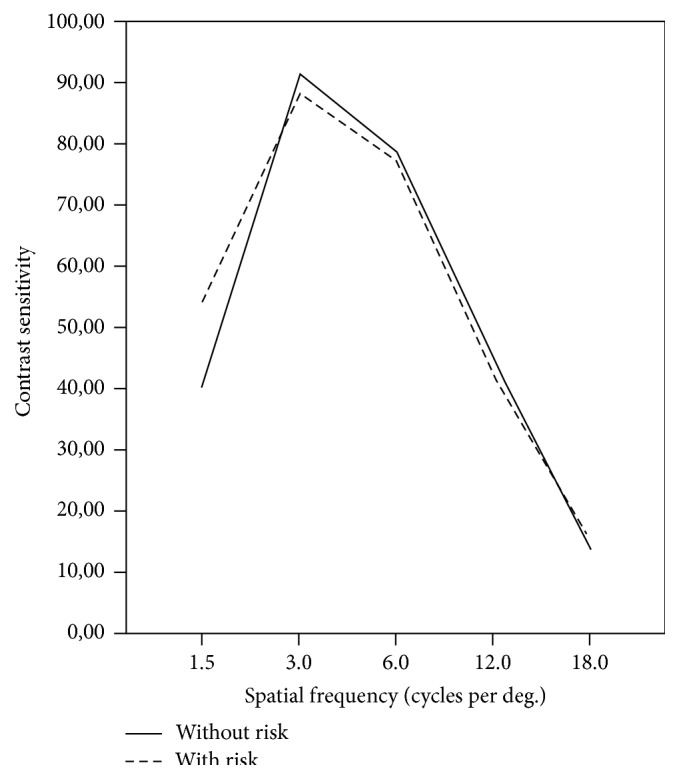
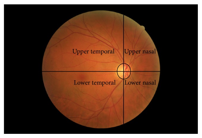
Purpose. Evaluate optimized fundus autofluorescence (FAF) imaging in early stages of diabetic retinopathy (DR) and relate findings with conventional colour fundus imaging and visual function in diabetic patients and control subjects. Materials and Methods. FAF and colour images were obtained using the CR-2 Plus digital nonmydriatic retinal camera in seven diabetic patients and thirteen control subjects. Visual-Functioning Questionnaire-25 (VFQ-25) and Diabetes Self-Management Questionnaire (DSMQ) were used to assess the quality of life and diabetes self-care. Contrast sensitivity function (CSF) was evaluated with the Vistech 6500 chart. Results. FAF and optimized-FAF imaging showed more retinal alterations related to DR than colour imaging. In diabetic patients, compatible signs with microaneurysms, capillary dilations, and haemorrhages were less numerous in colour imaging than optimized-FAF and FAF imaging in areas analysed. Control subjects at risk of developing DM showed more retinal pigment epithelium defects than those without risk in all retinal areas. Significant differences were not found in VFQ-25 and CSF between diabetic patients and control subjects. Conclusions. FAF and optimized-FAF imaging showed significant alterations related to DR not observed in colour imaging. FAF and optimized-FAF images could be a useful complementary tool for detecting early alterations associated with the development and progression of DR.
目的。评估优化后的眼底自发荧光(FAF)成像在糖尿病视网膜病变(DR)早期阶段的情况,并将结果与糖尿病患者和对照受试者的传统彩色眼底成像及视觉功能相关联。材料与方法。使用CR - 2 Plus数字免散瞳视网膜相机对7例糖尿病患者和13例对照受试者获取FAF和彩色图像。采用视觉功能问卷 - 25(VFQ - 25)和糖尿病自我管理问卷(DSMQ)评估生活质量和糖尿病自我护理情况。使用Vistech 6500图表评估对比敏感度功能(CSF)。结果。与彩色成像相比,FAF和优化后的FAF成像显示出更多与DR相关的视网膜改变。在糖尿病患者中,在分析区域内,彩色成像中与微动脉瘤、毛细血管扩张和出血相符的体征比优化后的FAF和FAF成像中更少。有患糖尿病风险的对照受试者在所有视网膜区域的视网膜色素上皮缺陷比无风险者更多。糖尿病患者和对照受试者在VFQ - 25和CSF方面未发现显著差异。结论。FAF和优化后的FAF成像显示出与DR相关的显著改变,而彩色成像中未观察到这些改变。FAF和优化后的FAF图像可能是检测与DR发生和发展相关的早期改变的有用补充工具。