Jiang Ruisheng, Zeng Xiangmin, Sun Shihang, Ma Zhijun, Wang Ximing
Diagnostic Room of Computer Tomography, Shandong Medical Imaging Research Institute, Shandong University, Jinan, Shandong, China (mainland).
Department of Computer Tomography and Magnetic Resonance Imaging, Weifang Medical College Affiliated Yidu Central Hospital, Qingzhou, Shandong, China (mainland).
Med Sci Monit. 2016 Apr 20;22:1318-28. doi: 10.12659/msm.895755.
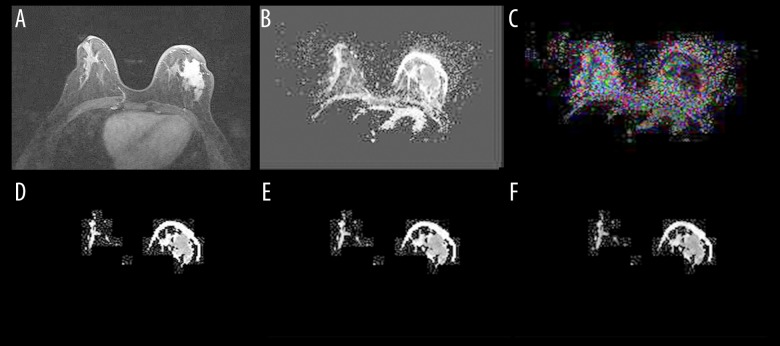
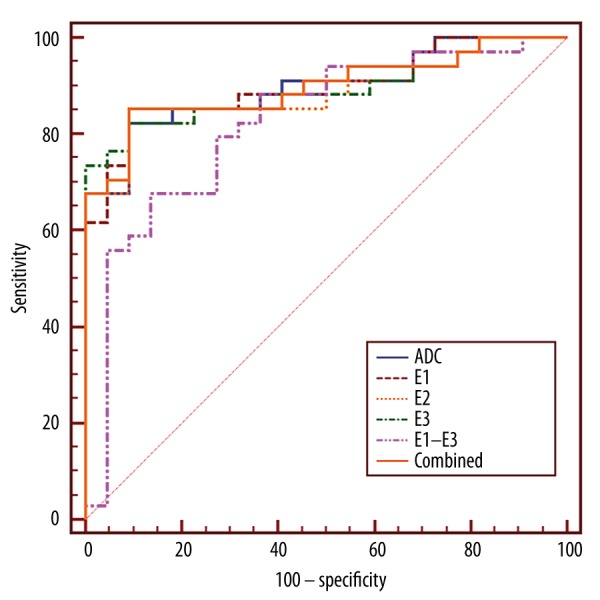
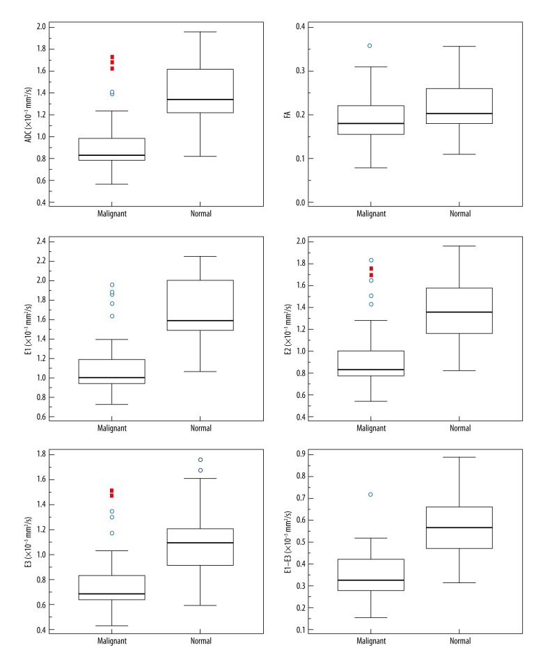
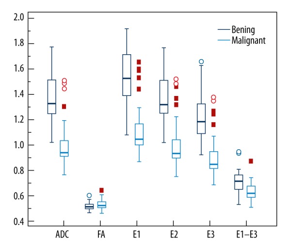
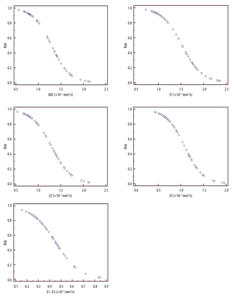
BACKGROUND The aim of this study was to investigate whether the anisotropy parameters are helpful in the detection and discrimination of breast cancers, and to determine its value in predicting the risk of cancers. MATERIAL AND METHODS There were 56 patients with 56 lesions (34 malignant, 22 benign) included in the study. DTI was performed in every patient and apparent diffusion coefficient (ADC), fractional anisotropy (FA), and eigenvalues E1, E2, and E3 were measured in every lesion and the normal breast tissue. RESULTS ADC, FA, and eigenvalues of E1, E2, E3, and E1-E3 in breast cancers were all significantly lower than in normal tissue (P<0.001 for all) with mean reduction of (32 ± 17)%, (24 ± 13)%, (33 ± 19)%, (32 ± 17)%, (31 ± 18)%, and (37 ± 20)% for ADC, FA, E1, E2, E3, and E1-E3, respectively. These parameters were also statistically lower in cancers than in benign lesions (P<0.01 for all), except FA (P>0.05). ADC, E1, E2, and E3 were very similar in discriminating breast cancers and benign lesions, with area under the curve (AUC) 0.885-0.898, sensitivity 73.5-85.3%, and specificity 90.9-100%. CONCLUSIONS ADC, E1, E2, E3, and E1-E3 are much lower in breast cancers than in normal tissue and benign lesions. The reduction of ADC, E1, E2, E3, and E1-E3 of a mass in the breast is highly associated with the risk of breast cancer, but the FA has no utility in breast cancer risk prediction.
背景 本研究的目的是调查各向异性参数是否有助于乳腺癌的检测和鉴别,并确定其在预测癌症风险中的价值。材料与方法 本研究纳入了56例患者的56个病灶(34个恶性,22个良性)。对每位患者进行扩散张量成像(DTI),并测量每个病灶及正常乳腺组织的表观扩散系数(ADC)、分数各向异性(FA)以及本征值E1、E2和E3。结果 乳腺癌的ADC、FA以及E1、E2、E3和E1-E3的本征值均显著低于正常组织(均P<0.001),ADC、FA、E1、E2、E3和E1-E3的平均降低幅度分别为(32±17)%、(24±13)%、(33±19)%、(32±17)%、(31±18)%和(37±20)%。除FA外(P>0.05),这些参数在癌症中也显著低于良性病灶(均P<0.01)。ADC、E1、E2和E3在鉴别乳腺癌和良性病灶方面非常相似,曲线下面积(AUC)为0.885-0.898,敏感性为73.5-85.3%,特异性为90.9-100%。结论 乳腺癌中的ADC、E1、E2、E3和E1-E3远低于正常组织和良性病灶。乳腺肿块的ADC、E1、E2、E3和E1-E3降低与乳腺癌风险高度相关,但FA在乳腺癌风险预测中无作用。