Moosavi Horieh, Darvishzadeh Fatemeh
Dental Materials Research Center, Department of Operative Dentistry, Mashhad Dental School, Mashhad University of Medical Sciences, Mashhad, Iran.
Open Dent J. 2016 Mar 25;10:69-78. doi: 10.2174/1874210616021000069. eCollection 2016.
This study investigated the effects of post bleaching treatments to prevent restaining and the change of enamel surface microhardness after dental bleaching in vitro.
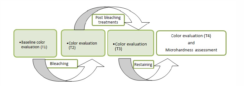
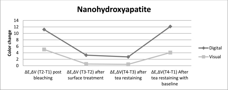
Sixty intact human incisor teeth were stained in tea solution and randomly assigned into four groups (n=15). Then samples were bleached for two weeks (8 hours daily) by 15% carbamide peroxide. Tooth color was determined both with a spectrophotometer and visually before bleaching (T1) and immediately after bleaching (T2). Next, it was applied in group 1 fluoride (Naf 2%) gel for 2 minutes, and in group 2 a fractional CO2 laser (10 mJ, 200 Hz, 10 s), and in group 3, nanohydroxyapatite gel for 2 minutes. The bleached teeth in group 4 remained untreated (control group). Then teeth placed in tea solution again. Color examinations were repeated after various post bleaching treatments (T3) and restaining with tea (T4) and color change values recorded. The microhardness was measured at the enamel surface of samples. Data was analyzed using ANOVA, Tukey HSD test and Dunnett T3 (α = 0.05).
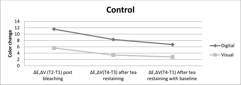
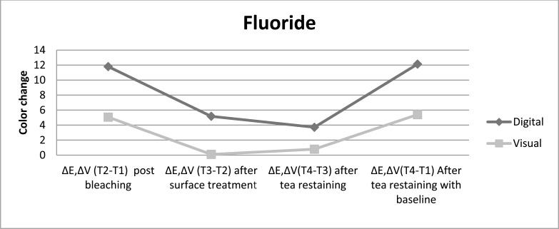
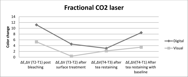
Directly after bleaching (ΔE T3-T2), the treatment with nanohydroxyapatite showed significantly the least color lapse in colorimetric evaluation. In experimental groups, the color change between T3 and T4 stages (ΔE T4-T3) was significantly lower than control group (P < 0.05). Different methods of enamel treatment caused a significant increase in surface microhardness compared to control group (P < 0.05).
Application of fluoride, fractional CO2 laser and nanohydroxyapatite as post bleaching treatments are suggested for prevention of stain absorption and increasing the hardening of bleached enamel.
本研究在体外调查了漂白后处理对预防再染色的效果以及牙齿漂白后釉质表面显微硬度的变化。
60颗完整的人切牙在茶溶液中染色,随机分为四组(n = 15)。然后用15%的过氧化脲对样本进行两周的漂白(每天8小时)。在漂白前(T1)和漂白后立即(T2)用分光光度计和肉眼测定牙齿颜色。接下来,第1组应用2%氟化钠(Naf)凝胶2分钟,第2组应用分次二氧化碳激光(10 mJ,200 Hz,10秒),第3组应用纳米羟基磷灰石凝胶2分钟。第4组的漂白牙齿不做处理(对照组)。然后将牙齿再次置于茶溶液中。在各种漂白后处理(T3)和用茶再染色(T4)后重复颜色检查并记录颜色变化值。在样本的釉质表面测量显微硬度。使用方差分析、Tukey HSD检验和Dunnett T3检验(α = 0.05)分析数据。
漂白后直接测量(ΔE T3 - T2),在比色评估中,纳米羟基磷灰石处理显示颜色变化明显最小。在实验组中,T3和T4阶段之间的颜色变化(ΔE T4 - T3)明显低于对照组(P < 0.05)。与对照组相比,不同的釉质处理方法导致表面显微硬度显著增加(P < 0.05)。
建议应用氟化物、分次二氧化碳激光和纳米羟基磷灰石作为漂白后处理,以预防污渍吸收并增加漂白釉质的硬度。