Pinto Maria J, Pedro Joana R, Costa Rui O, Almeida Ramiro D
Center for Neuroscience and Cell Biology (CNC), University of CoimbraCoimbra, Portugal; PhD Programme in Experimental Biology and Biomedicine (PDBEB), Center for Neuroscience and Cell Biology, University of CoimbraCoimbra, Portugal.
Center for Neuroscience and Cell Biology (CNC), University of Coimbra Coimbra, Portugal.
Front Mol Neurosci. 2016 Jun 10;9:43. doi: 10.3389/fnmol.2016.00043. eCollection 2016.
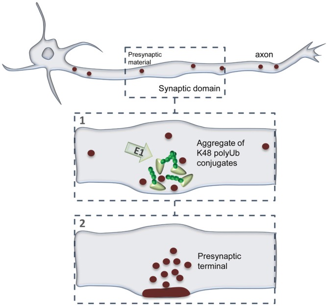
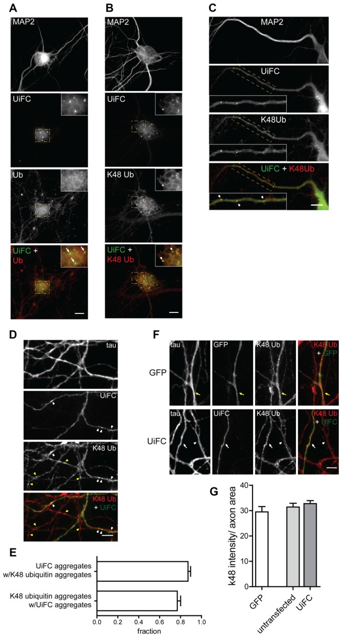
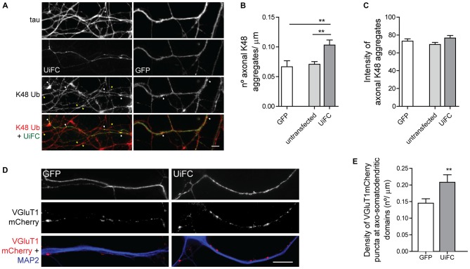
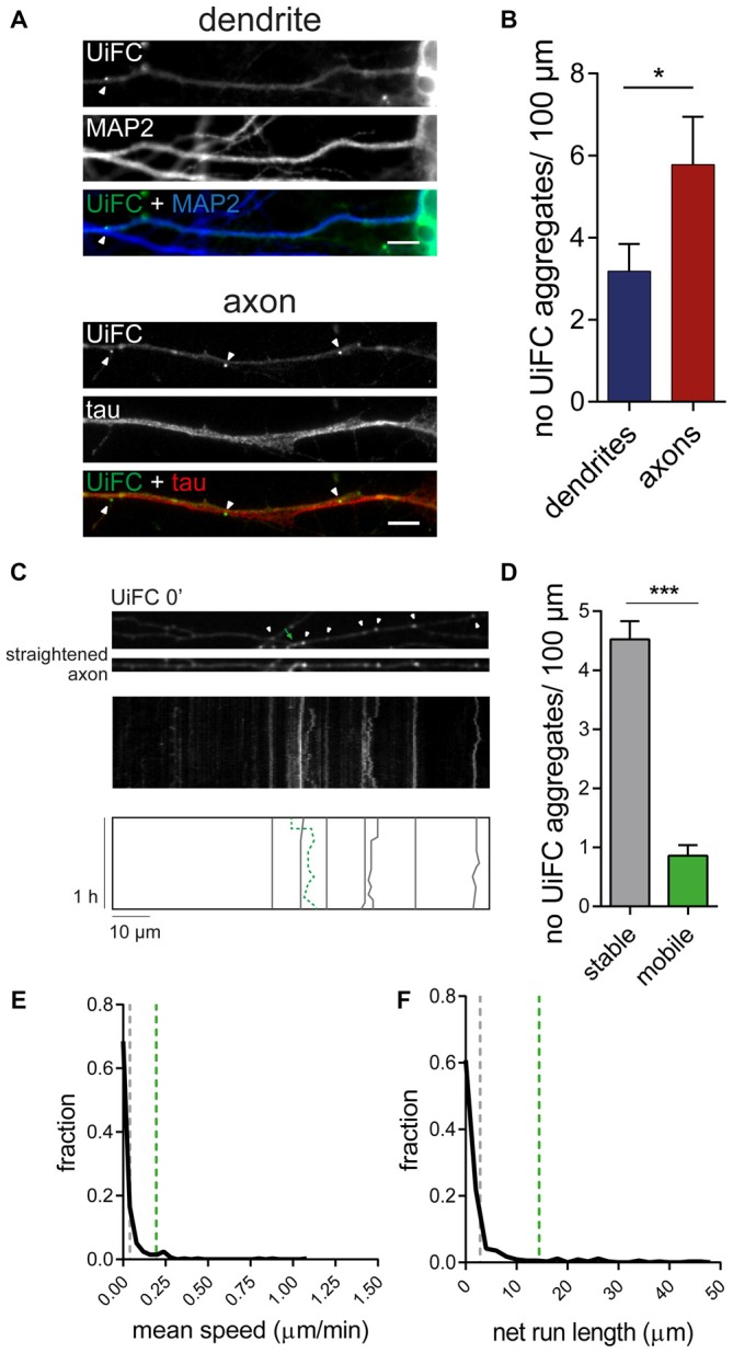
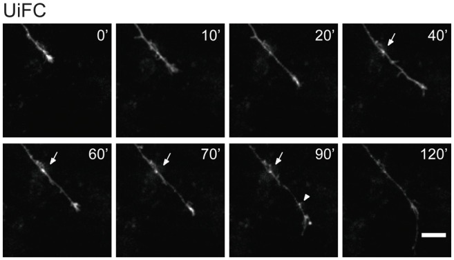
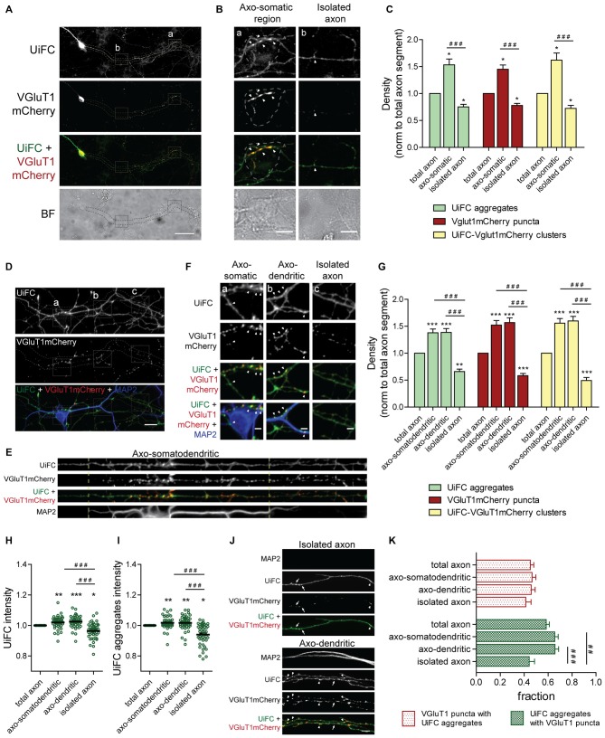
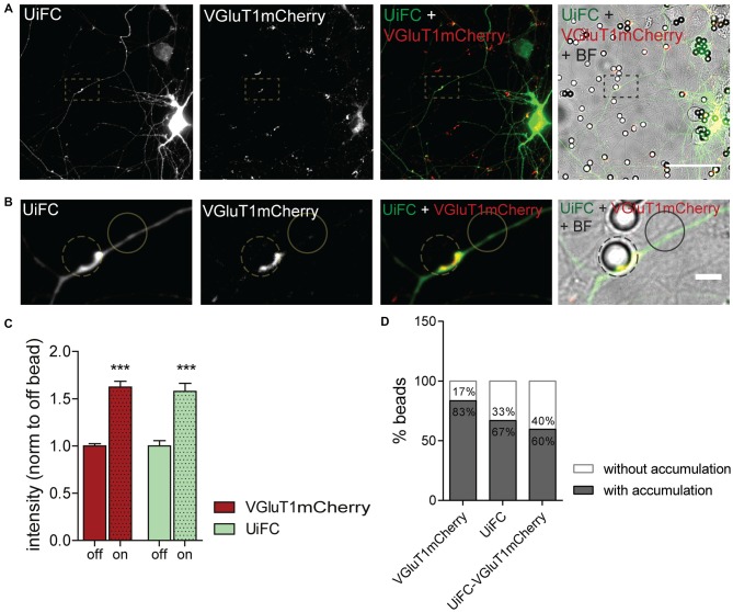
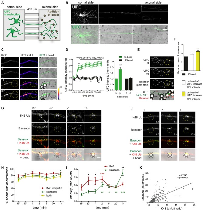
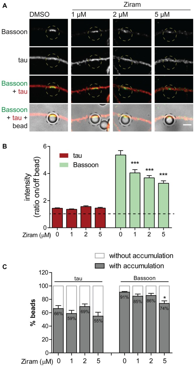
In recent years, signaling through ubiquitin has been shown to be of great importance for normal brain development. Indeed, fluctuations in ubiquitin levels and spontaneous mutations in (de)ubiquitination enzymes greatly perturb synapse formation and neuronal transmission. In the brain, expression of lysine (K) 48-linked ubiquitin chains is higher at a developmental stage coincident with synaptogenesis. Nevertheless, no studies have so far delved into the involvement of this type of polyubiquitin chains in synapse formation. We have recently proposed a role for polyubiquitinated conjugates as triggering signals for presynaptic assembly. Herein, we aimed at characterizing the axonal distribution of K48 polyubiquitin and its dynamics throughout the course of presynaptic formation. To accomplish so, we used an ubiquitination-induced fluorescence complementation (UiFC) strategy for the visualization of K48 polyubiquitin in live hippocampal neurons. We first validated its use in neurons by analyzing changing levels of polyubiquitin. UiFC signal is diffusely distributed with distinct aggregates in somas, dendrites and axons, which perfectly colocalize with staining for a K48-specific antibody. Axonal UiFC aggregates are relatively stable and new aggregates are formed as an axon grows. Approximately 65% of UiFC aggregates colocalize with synaptic vesicle clusters and they preferentially appear in the axonal domains of axo-somatodendritic synapses when compared to isolated axons. We then evaluated axonal accumulation of K48 ubiquitinated signals in bead-induced synapses. We observed rapid accumulation of UiFC signal and endogenous K48 ubiquitin at the sites of newly formed presynapses. Lastly, we show by means of a microfluidic platform, for the isolation of axons, that presynaptic clustering on beads is dependent on E1-mediated ubiquitination at the axonal level. Altogether, these results indicate that enrichment of K48 polyubiquitin at the site of nascent presynaptic terminals is an important axon-intrinsic event for presynaptic differentiation.
近年来,泛素信号通路已被证明对正常脑发育至关重要。事实上,泛素水平的波动以及(去)泛素化酶的自发突变会极大地扰乱突触形成和神经元传递。在大脑中,与突触发生同时的发育阶段,赖氨酸(K)48连接的泛素链表达较高。然而,迄今为止尚无研究深入探讨这类多聚泛素链在突触形成中的作用。我们最近提出多聚泛素化偶联物作为突触前组装触发信号的作用。在此,我们旨在表征K48多聚泛素在轴突中的分布及其在突触前形成过程中的动态变化。为实现这一目标,我们采用泛素化诱导荧光互补(UiFC)策略来可视化活海马神经元中的K48多聚泛素。我们首先通过分析多聚泛素水平的变化来验证其在神经元中的应用。UiFC信号在胞体、树突和轴突中呈弥散分布并伴有明显聚集,这些聚集与K48特异性抗体染色完美共定位。轴突中的UiFC聚集物相对稳定,并且随着轴突生长会形成新的聚集物。大约65%的UiFC聚集物与突触小泡簇共定位,与孤立轴突相比,它们更优先出现在轴 - 体树突突触的轴突区域。然后我们评估了在珠子诱导的突触中K48泛素化信号的轴突积累。我们观察到UiFC信号和内源性K48泛素在新形成的突触前位点迅速积累。最后,我们通过微流控平台分离轴突表明,珠子上的突触前聚集依赖于轴突水平的E1介导的泛素化。总之,这些结果表明新生突触前终末位点处K48多聚泛素的富集是突触前分化的一个重要轴突内在事件。