Iwahashi Hiroki, Yoshimura Noriko, Hashizume Hiroshi, Yamada Hiroshi, Oka Hiroyuki, Matsudaira Ko, Shinto Kazunori, Ishimoto Yuyu, Nagata Keiji, Teraguchi Masatoshi, Kagotani Ryohei, Muraki Shigeyuki, Akune Toru, Tanaka Sakae, Kawaguchi Hiroshi, Nakamura Kozo, Minamide Akihito, Nakagawa Yukihiro, Yoshida Munehito
Department of Orthopaedic Surgery, Wakayama Medical University, 811-1 Kimiidera, Wakayama City, Wakayama 641-8510, Japan.
Department of Joint Disease Research, 22nd Century Medical and Research Center, Faculty of Medicine, The University of Tokyo, 7-3-1 Hongo, Bunkyo-ku, Tokyo 113-8655, Japan.
PLoS One. 2016 Aug 3;11(8):e0160002. doi: 10.1371/journal.pone.0160002. eCollection 2016.
The purpose of this study was to evaluate the relations between the degree of encroachment, measured as the cross-sectional area of the dural sac, and low back pain in a large population.
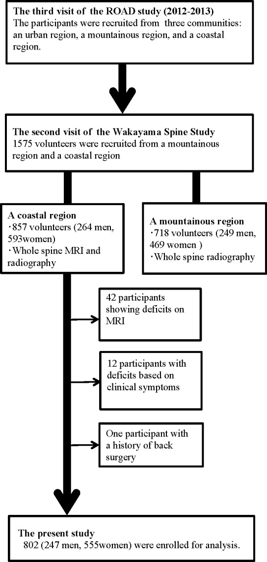
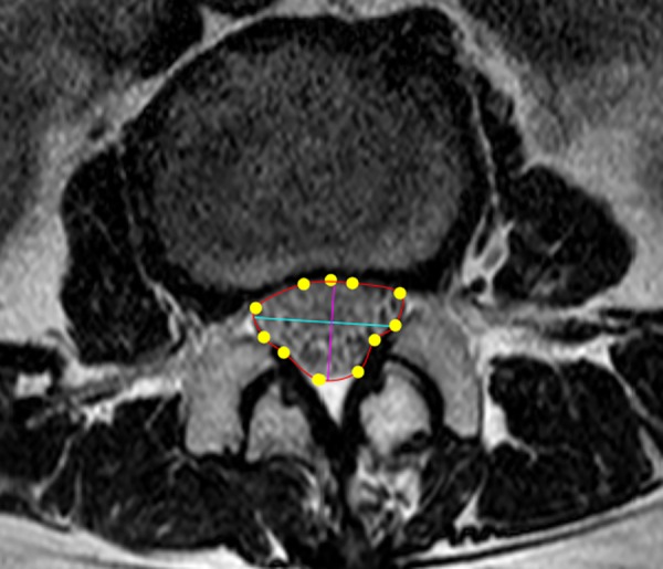
In this cross-sectional study, data from 802 participants (247 men, 555 women; mean age, 63.5 years) were analyzed. The measurement of the cross-sectional area of the dural sac from the level of L1/2 to L4/5 was taken using axial T2-weighted images. The minimum cross-sectional area was defined as the cross-sectional area of the dural sac at the most constricted level in the examined spine. Participants were divided into three groups according to minimum cross-sectional area measurement quartiles (less than the first quartile, between the first and third quartiles, and greater than the third quartile). A multivariate logistic regression analysis was used to estimate the association between the minimum cross-sectional area and the prevalence of low back pain.
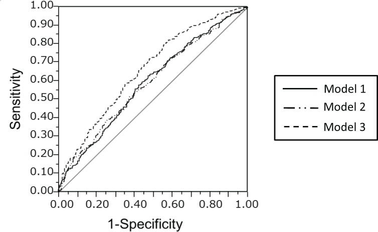
The mean minimum cross-sectional area was 117.3 mm2 (men: 114.4 mm2; women: 118.6 mm2). A logistic regression analysis adjusted for age, sex, body mass index, and other confounding factors, including disc degeneration, showed that a narrow minimum cross-sectional area (smaller than the first quartile) was significantly associated with low back pain (odds ratio, 1.78; 95% confidence interval, 1.13-2.80 compared to the wide minimum cross-sectional area group: minimum cross-sectional area greater than the third quartile measured).
This study showed that a narrow dural sac cross-sectional area was significantly associated with the presence of low back pain after adjustment for age, sex, and body mass index. Further investigations that include additional radiographic findings and psychological factors will continue to elucidate the causes of low back pain.
本研究旨在评估以硬脊膜囊横截面积衡量的受压程度与大量人群中腰痛之间的关系。
在这项横断面研究中,分析了802名参与者(247名男性,555名女性;平均年龄63.5岁)的数据。使用轴向T2加权图像测量从L1/2至L4/5水平的硬脊膜囊横截面积。最小横截面积定义为所检查脊柱中最狭窄水平处硬脊膜囊的横截面积。参与者根据最小横截面积测量四分位数分为三组(小于第一四分位数、在第一和第三四分位数之间、大于第三四分位数)。采用多因素逻辑回归分析来估计最小横截面积与腰痛患病率之间的关联。
平均最小横截面积为117.3平方毫米(男性:114.4平方毫米;女性:118.6平方毫米)。经年龄、性别、体重指数以及包括椎间盘退变在内的其他混杂因素校正后的逻辑回归分析表明,最小横截面积狭窄(小于第一四分位数)与腰痛显著相关(比值比,1.78;95%置信区间,与最小横截面积宽的组相比:测量的最小横截面积大于第三四分位数,为1.13 - 2.80)。
本研究表明,在对年龄、性别和体重指数进行校正后,硬脊膜囊横截面积狭窄与腰痛的存在显著相关。包括更多影像学发现和心理因素的进一步研究将继续阐明腰痛的原因。