Valsky Dan, Marmor-Levin Odeya, Deffains Marc, Eitan Renana, Blackwell Kim T, Bergman Hagai, Israel Zvi
The Edmond and Lily Safra Center for Brain Research (ELSC), The Hebrew University, Jerusalem, Israel.
Department of Medical Neurobiology (Physiology), Institute of Medical Research - Israel-Canada (IMRIC), The Hebrew University-Hadassah Medical School, Jerusalem, Israel.
Mov Disord. 2017 Jan;32(1):70-79. doi: 10.1002/mds.26806. Epub 2016 Oct 6.
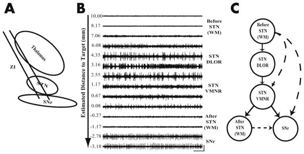
Microelectrode recordings along preplanned trajectories are often used for accurate definition of the subthalamic nucleus (STN) borders during deep brain stimulation (DBS) surgery for Parkinson's disease. Usually, the demarcation of the STN borders is performed manually by a neurophysiologist. The exact detection of the borders is difficult, especially detecting the transition between the STN and the substantia nigra pars reticulata. Consequently, demarcation may be inaccurate, leading to suboptimal location of the DBS lead and inadequate clinical outcomes.
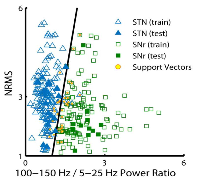
We present machine-learning classification procedures that use microelectrode recording power spectra and allow for real-time, high-accuracy discrimination between the STN and substantia nigra pars reticulata.
A support vector machine procedure was tested on microelectrode recordings from 58 trajectories that included both STN and substantia nigra pars reticulata that achieved a 97.6% consistency with human expert classification (evaluated by 10-fold cross-validation). We used the same data set as a training set to find the optimal parameters for a hidden Markov model using both microelectrode recording features and trajectory history to enable real-time classification of the ventral STN border (STN exit). Seventy-three additional trajectories were used to test the reliability of the learned statistical model in identifying the exit from the STN. The hidden Markov model procedure identified the STN exit with an error of 0.04 ± 0.18 mm and detection reliability (error < 1 mm) of 94%.
The results indicate that robust, accurate, and automatic real-time electrophysiological detection of the ventral STN border is feasible. © 2016 International Parkinson and Movement Disorder Society.
在帕金森病的脑深部电刺激(DBS)手术中,沿预先规划的轨迹进行微电极记录常用于精确界定丘脑底核(STN)边界。通常,STN边界的划分由神经生理学家手动完成。精确检测边界很困难,尤其是检测STN与黑质网状部之间的过渡区域。因此,边界划分可能不准确,导致DBS电极位置不理想,临床效果不佳。
我们提出了利用微电极记录功率谱的机器学习分类程序,可实时、高精度地区分STN和黑质网状部。
在包含STN和黑质网状部的58条轨迹的微电极记录上测试了支持向量机程序,其与人类专家分类的一致性达到97.6%(通过10折交叉验证评估)。我们使用相同的数据集作为训练集,以找到使用微电极记录特征和轨迹历史的隐马尔可夫模型的最佳参数,从而实现对腹侧STN边界(STN出口)的实时分类。另外73条轨迹用于测试所学统计模型识别STN出口的可靠性。隐马尔可夫模型程序识别STN出口的误差为0.04±0.18毫米,检测可靠性(误差<1毫米)为94%。
结果表明,对腹侧STN边界进行稳健、准确且自动的实时电生理检测是可行的。©2016国际帕金森病和运动障碍协会。