Page Shyanne, Munsell Alli, Al-Ahmad Abraham J
Department of Pharmaceutical Sciences, School of Pharmacy, Texas Tech University Health Sciences Center, 1300 South Coulter Street, Amarillo, TX, USA.
Fluids Barriers CNS. 2016 Oct 11;13(1):16. doi: 10.1186/s12987-016-0042-1.
Cerebral hypoxia/ischemia (H/I) is an important stress factor involved in the disruption of the blood-brain barrier (BBB) following stroke injury, yet the cellular and molecular mechanisms on how the human BBB responds to such injury remains unclear. In this study, we investigated the cellular response of the human BBB to chemical and environmental H/I in vitro.
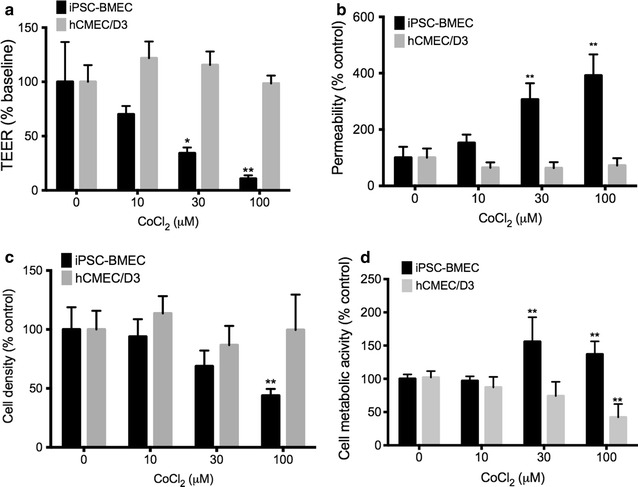
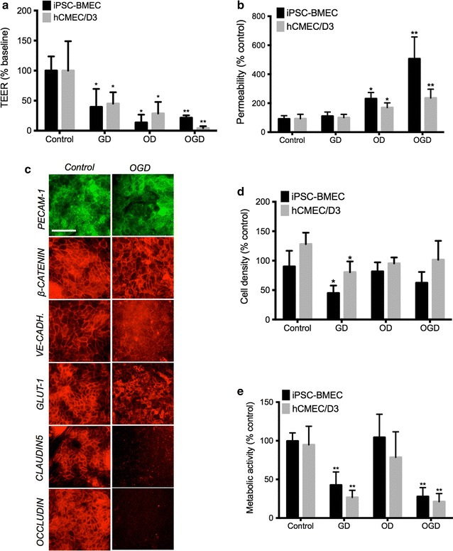
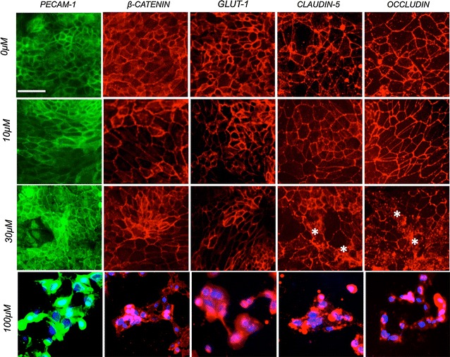
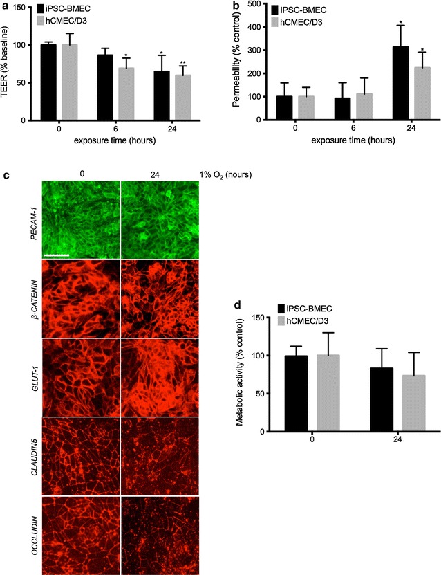
In this study, we used immortalized hCMEC/D3 and IMR90 stem-cell derived human brain microvascular endothelial cell lines (IMR90-derived BMECs). Hypoxic stress was achieved by exposure to cobalt chloride (CoCl) or by exposure to 1 % hypoxia and oxygen/glucose deprivation (OGD) was used to model ischemic injury. We assessed barrier function using both transendothelial electrical resistance (TEER) and sodium fluorescein permeability. Changes in cell junction integrity were assessed by immunocytochemistry and cell viability was assessed by trypan-blue exclusion and by MTS assays. Statistical analysis was performed using one-way analysis of variance (ANOVA).
CoCl selectively disrupted the barrier function in IMR90-derived BMECs but not in hCMEC/D3 monolayers and cytotoxic effects did not drive such disruption. In addition, hypoxia/OGD stress significantly disrupted the barrier function by selectively disrupting tight junctions (TJs) complexes. In addition, we noted an uncoupling between cell metabolic activity and barrier integrity.
In this study, we demonstrated the ability of IMR90-derived BMECs to respond to hypoxic/ischemic injury triggered by both chemical and environmental stress by showing a disruption of the barrier function. Such disruption was selectively targeting TJ complexes and was not driven by cellular apoptosis. In conclusion, this study suggests the suitability of stem cell-derived human BMECs monolayers as a model of cerebral hypoxia/ischemia in vitro.
脑缺氧/缺血(H/I)是中风损伤后血脑屏障(BBB)破坏所涉及的一个重要应激因素,但人类血脑屏障如何应对此类损伤的细胞和分子机制仍不清楚。在本研究中,我们在体外研究了人类血脑屏障对化学性和环境性H/I的细胞反应。
在本研究中,我们使用了永生化的hCMEC/D3和IMR90干细胞衍生的人脑微血管内皮细胞系(IMR90衍生的BMECs)。通过暴露于氯化钴(CoCl)或暴露于1%低氧来实现缺氧应激,并用氧/葡萄糖剥夺(OGD)来模拟缺血性损伤。我们使用跨内皮电阻(TEER)和荧光素钠通透性来评估屏障功能。通过免疫细胞化学评估细胞连接完整性的变化,通过台盼蓝排斥法和MTS试验评估细胞活力。使用单因素方差分析(ANOVA)进行统计分析。
CoCl选择性破坏了IMR90衍生的BMECs中的屏障功能,但未破坏hCMEC/D3单层细胞的屏障功能,细胞毒性作用并未导致这种破坏。此外,缺氧/OGD应激通过选择性破坏紧密连接(TJ)复合物显著破坏了屏障功能。此外,我们注意到细胞代谢活性与屏障完整性之间存在解偶联。
在本研究中,我们通过显示屏障功能的破坏,证明了IMR90衍生的BMECs对化学和环境应激引发的缺氧/缺血性损伤的反应能力。这种破坏选择性地靶向TJ复合物,并非由细胞凋亡驱动。总之,本研究表明干细胞衍生的人BMECs单层细胞适合作为体外脑缺氧/缺血的模型。