Park Jae-Hyeong, Lee Ju-Hee, Lee Sang Yeub, Choi Jin-Oh, Shin Mi-Seung, Kim Mi-Jeong, Jung Hae Ok, Park Jeong Rang, Sohn Il Suk, Kim Hyungseop, Park Seong-Mi, Yoo Nam Jin, Choi Jung Hyun, Kim Hyung-Kwan, Cho Goo-Yeong, Lee Mi-Rae, Park Jin-Sun, Shim Chi Young, Kim Dae-Hee, Shin Dae-Hee, Shin Gil Ja, Shin Sung Hee, Kim Kye Hun, Kim Woo-Shik, Park Seung Woo
Division of Cardiology, Department of Internal Medicine, Chungnam National University Hospital, Chungnam National University School of Medicine, Daejeon, Korea.
Division of Cardiology, Department of Internal Medicine, Chungbuk National University School of Medicine, Cheongju, Korea.
J Cardiovasc Ultrasound. 2016 Dec;24(4):285-293. doi: 10.4250/jcu.2016.24.4.285. Epub 2016 Dec 28.
It is important to understand the distribution of 2-dimensional strain values in normal population. We performed a multicenter trial to measure normal echocardiographic values in the Korean population.
This was a substudy of the Normal echOcardiogRaphic Measurements in KoreAn popuLation (NORMAL) study. Echocardiographic specialists measured frequently used echocardiographic indices in healthy people according to a standardized method at 23 different university hospitals. The strain values were analyzed from digitally stored images.
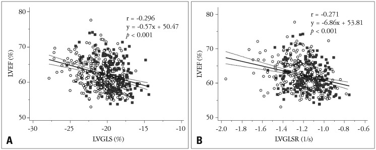
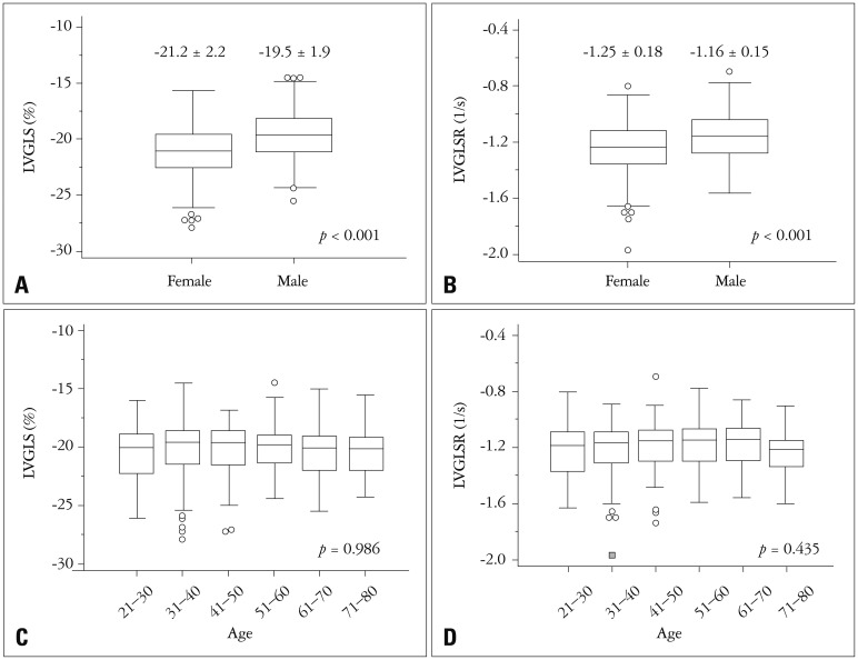
Of a total of 1003 healthy participants in NORMAL study, 2-dimensional strain values were measured in 501 subjects (265 females, mean age 47 ± 15 years old) with echocardiographic images only by GE echocardiographic machines. Interventricular septal thickness, left ventricular (LV) posterior wall thickness, systolic and diastolic LV dimensions, and LV ejection fraction were 7.5 ± 1.0 mm, 7.4 ± 1.0 mm, 29.9 ± 2.8 mm, 48.9 ± 3.6 mm, and 62 ± 4%, respectively. LV longitudinal systolic strain (LS) values of apical 4-chamber (A4C) view, apical 3-chamber (A3C) view, apical 2-chamber (A2C) view, and LV global LS (LVGLS) were -20.1 ± 2.3, -19.9 ± 2.7, -21.2 ± 2.6, and -20.4 ± 2.2%, respectively. LV longitudinal systolic strain rate (LVLSR) values of the A4C view, A3C view, A2C view, and LV global LSR (LVGLSR) were -1.18 ± 0.18, -1.20 ± 0.21, -1.25 ± 0.21, and -1.21 ± 0.21, respectively. Females had lower LVGLS (-21.2 ± 2.2% vs. -19.5 ± 1.9%, < 0.001) and LVGLSR (-1.25 ± 0.18 vs. -1.17 ± 0.15, < 0.001) values than males.
We measured LV longitudinal strain and strain rate values in the normal Korean population. Since considerable gender differences were observed, normal echocardiographic cutoff values should be differentially applied based on sex.
了解正常人群二维应变值的分布很重要。我们进行了一项多中心试验,以测量韩国人群的正常超声心动图值。
这是韩国人群正常超声心动图测量(NORMAL)研究的一项子研究。超声心动图专家在23家不同的大学医院,根据标准化方法对健康人常用的超声心动图指标进行测量。应变值从数字存储的图像中分析得出。
在NORMAL研究的1003名健康参与者中,仅通过GE超声心动图机器对501名受试者(265名女性,平均年龄47±15岁)进行了二维应变值测量。室间隔厚度、左心室(LV)后壁厚度、左心室收缩和舒张尺寸以及左心室射血分数分别为7.5±1.0毫米、7.4±1.0毫米、29.9±2.8毫米、48.9±3.6毫米和62±4%。心尖四腔心(A4C)视图、心尖三腔心(A3C)视图、心尖二腔心(A2C)视图的左心室纵向收缩应变(LS)值以及左心室整体LS(LVGLS)分别为-20.1±2.3%、-19.9±2.7%、-21.2±2.6%和-20.4±2.2%。A4C视图、A3C视图、A2C视图的左心室纵向收缩应变率(LVLSR)值以及左心室整体LSR(LVGLSR)分别为-1.18±0.18、-1.20±0.21、-1.25±0.21和-1.21±0.21。女性的LVGLS(-21.2±2.2%对-19.5±1.9%,<0.001)和LVGLSR(-1.25±0.18对-1.17±0.15,<0.001)值低于男性。
我们测量了韩国正常人群的左心室纵向应变和应变率值。由于观察到显著的性别差异,正常超声心动图临界值应根据性别差异应用。