Sobotta Katharina, Bonkowski Katharina, Liebler-Tenorio Elisabeth, Germon Pierre, Rainard Pascal, Hambruch Nina, Pfarrer Christiane, Jacobsen Ilse D, Menge Christian
Institute of Molecular Pathogenesis, Friedrich-Loeffler-Institut (FLI), Naumburger Strasse 96a, 07743, Jena, Germany.
ISP, INRA, Université Tours, UMR 1282, 37380, Nouzilly, France.
Vet Res. 2017 Apr 12;48(1):23. doi: 10.1186/s13567-017-0430-9.
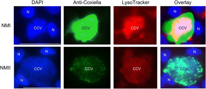
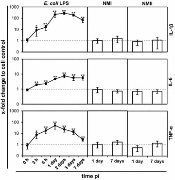
Ruminants are the main source of human infections with the obligate intracellular bacterium Coxiella (C.) burnetii. Infected animals shed high numbers of C. burnetii by milk, feces, and birth products. In goats, shedding by the latter route coincides with C. burnetii replication in epithelial (trophoblast) cells of the placenta, which led us to hypothesize that epithelial cells are generally implicated in replication and shedding of C. burnetii. We therefore aimed at analyzing the interactions of C. burnetii with epithelial cells of the bovine host (1) at the entry site (lung epithelium) which govern host immune responses and (2) in epithelial cells of gut, udder and placenta decisive for the quantity of pathogen excretion. Epithelial cell lines [PS (udder), FKD-R 971 (small intestine), BCEC (maternal placenta), F3 (fetal placenta), BEL-26 (lung)] were inoculated with C. burnetii strains Nine Mile I (NMI) and NMII at different cultivation conditions. The cell lines exhibited different permissiveness for C. burnetii. While maintaining cell viability, udder cells allowed the highest replication rates with formation of large cell-filling Coxiella containing vacuoles. Intestinal cells showed an enhanced susceptibility to invasion but supported C. burnetii replication only at intermediate levels. Lung and placental cells also internalized the bacteria but in strikingly smaller numbers. In any of the epithelial cells, both Coxiella strains failed to trigger a substantial IL-1β, IL-6 and TNF-α response. Epithelial cells, with mammary epithelial cells in particular, may therefore serve as a niche for C. burnetii replication in vivo without alerting the host's immune response.
反刍动物是人类感染专性细胞内细菌伯氏考克斯体(Coxiella (C.) burnetii)的主要来源。受感染的动物通过乳汁、粪便和分娩产物排出大量的伯氏考克斯体。在山羊中,通过后一种途径排出伯氏考克斯体与该菌在胎盘上皮(滋养层)细胞中的复制同时发生,这使我们推测上皮细胞通常与伯氏考克斯体的复制和排出有关。因此,我们旨在分析伯氏考克斯体与牛宿主上皮细胞的相互作用:(1)在控制宿主免疫反应的进入部位(肺上皮);(2)在对病原体排泄量起决定性作用的肠道、乳腺和胎盘的上皮细胞中。在不同培养条件下,用伯氏考克斯体菌株九里I(Nine Mile I,NMI)和九里II(NMII)接种上皮细胞系[PS(乳腺)、FKD-R 971(小肠)、BCEC(母体胎盘)、F3(胎儿胎盘)、BEL-26(肺)]。这些细胞系对伯氏考克斯体表现出不同的易感性。在维持细胞活力的同时,乳腺细胞允许最高的复制率,并形成充满细胞的、含有伯氏考克斯体的大液泡。肠道细胞对入侵的敏感性增强,但仅在中等水平支持伯氏考克斯体的复制。肺和胎盘细胞也摄取了细菌,但数量明显较少。在任何一种上皮细胞中,两种考克斯体菌株均未能引发显著的IL-1β、IL-6和TNF-α反应。因此,上皮细胞,尤其是乳腺上皮细胞,可能是伯氏考克斯体在体内复制的场所,而不会引发宿主的免疫反应。