• 文献检索
  • 文档翻译
  • 深度研究
  • 学术资讯
  • Suppr Zotero 插件Zotero 插件
  • 邀请有礼
  • 套餐&价格
  • 历史记录
应用&插件
Suppr Zotero 插件Zotero 插件浏览器插件Mac 客户端Windows 客户端微信小程序
定价
高级版会员购买积分包购买API积分包
服务
文献检索文档翻译深度研究API 文档MCP 服务
关于我们
关于 Suppr公司介绍联系我们用户协议隐私条款
关注我们

Suppr 超能文献

核心技术专利:CN118964589B侵权必究
粤ICP备2023148730 号-1Suppr @ 2026

文献检索

告别复杂PubMed语法,用中文像聊天一样搜索,搜遍4000万医学文献。AI智能推荐,让科研检索更轻松。

立即免费搜索

文件翻译

保留排版,准确专业,支持PDF/Word/PPT等文件格式,支持 12+语言互译。

免费翻译文档

深度研究

AI帮你快速写综述,25分钟生成高质量综述,智能提取关键信息,辅助科研写作。

立即免费体验

果蝇大脑咽下神经节的结构与发育。I. 节段结构、分区及谱系解剖学

Structure and development of the subesophageal zone of the Drosophila brain. I. Segmental architecture, compartmentalization, and lineage anatomy.

作者信息

Hartenstein Volker, Omoto Jaison J, Ngo Kathy T, Wong Darren, Kuert Philipp A, Reichert Heinrich, Lovick Jennifer K, Younossi-Hartenstein Amelia

机构信息

Department of Molecular Cell and Developmental Biology, University of California Los Angeles, Los Angeles, California.

Biozentrum, University of Basel, Basel, Switzerland.

出版信息

J Comp Neurol. 2018 Jan 1;526(1):6-32. doi: 10.1002/cne.24287. Epub 2017 Aug 10.

DOI:10.1002/cne.24287
PMID:28730682
原文链接:https://pmc.ncbi.nlm.nih.gov/articles/PMC5963519/
Abstract

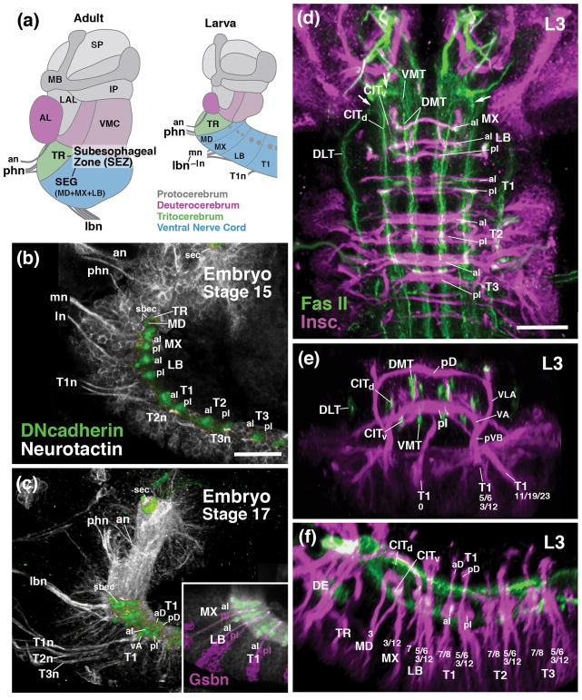
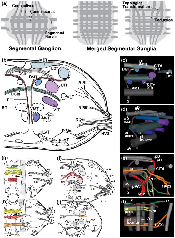
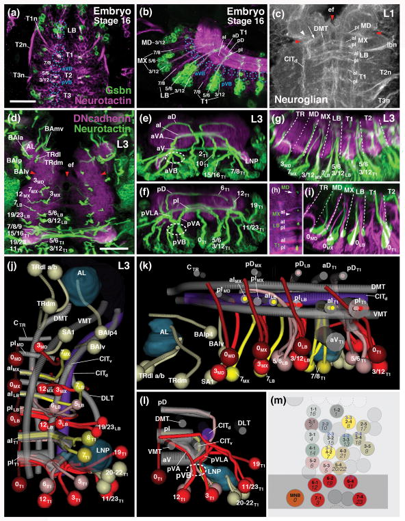
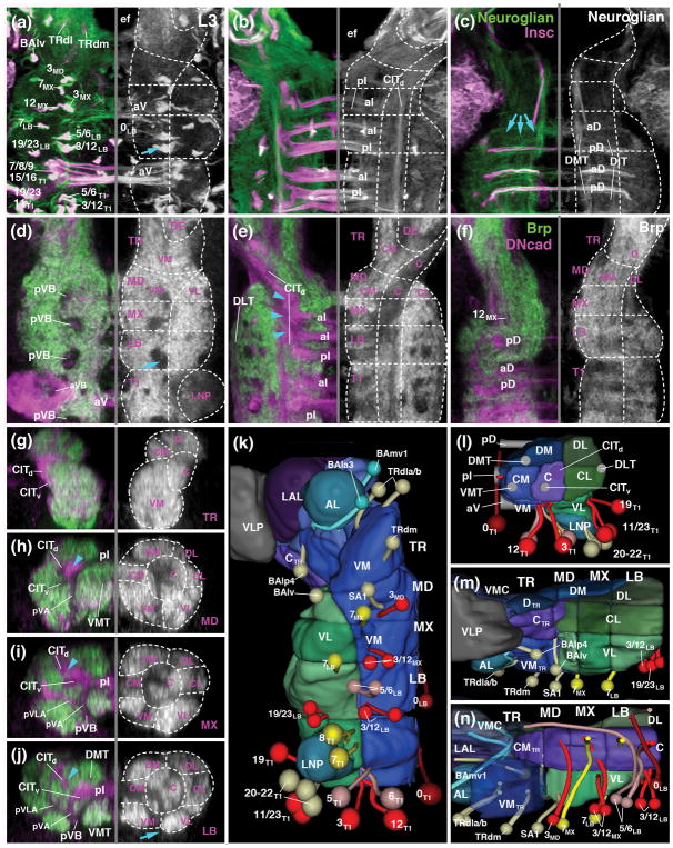
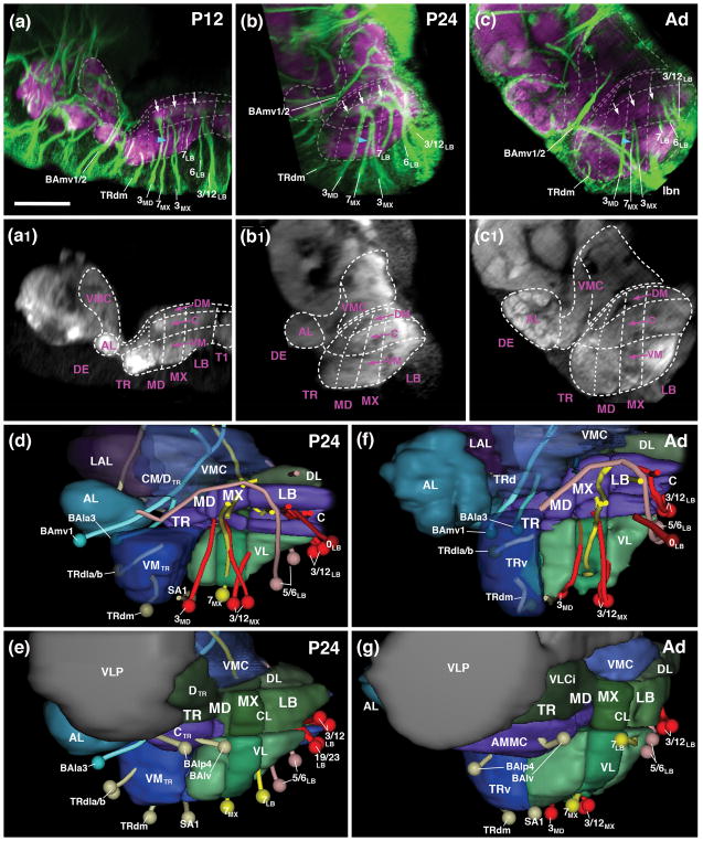
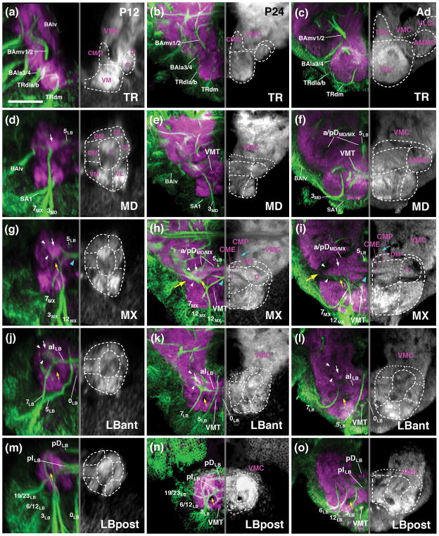
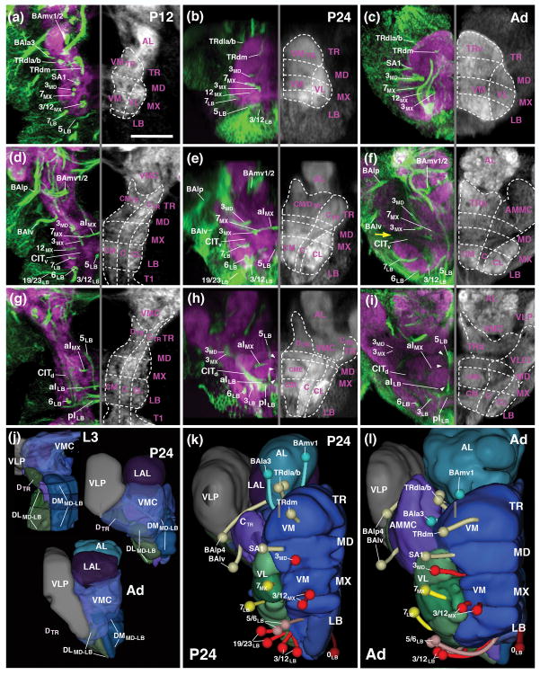
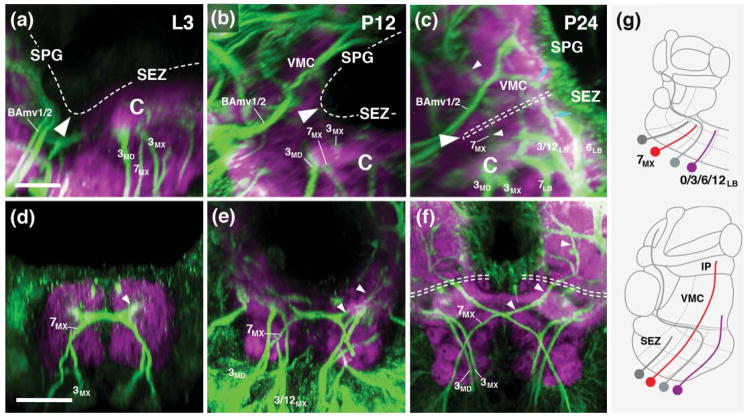
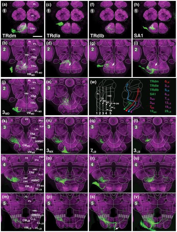
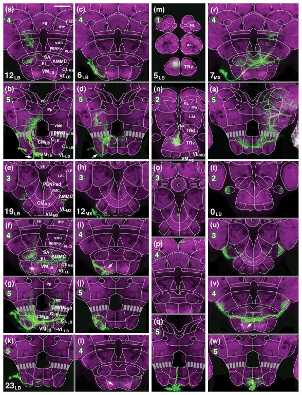
The subesophageal zone (SEZ) of the Drosophila brain houses the circuitry underlying feeding behavior and is involved in many other aspects of sensory processing and locomotor control. Formed by the merging of four neuromeres, the internal architecture of the SEZ can be best understood by identifying segmentally reiterated landmarks emerging in the embryo and larva, and following the gradual changes by which these landmarks become integrated into the mature SEZ during metamorphosis. In previous works, the system of longitudinal fibers (connectives) and transverse axons (commissures) has been used as a scaffold that provides internal landmarks for the neuromeres of the larval ventral nerve cord. We have extended the analysis of this scaffold to the SEZ and, in addition, reconstructed the tracts formed by lineages and nerves in relationship to the connectives and commissures. As a result, we establish reliable criteria that define boundaries between the four neuromeres (tritocerebrum, mandibular neuromere, maxillary neuromere, labial neuromere) of the SEZ at all stages of development. Fascicles and lineage tracts also demarcate seven columnar neuropil domains (ventromedial, ventro-lateral, centromedial, central, centrolateral, dorsomedial, dorsolateral) identifiable throughout development. These anatomical subdivisions, presented in the form of an atlas including confocal sections and 3D digital models for the larval, pupal and adult stage, allowed us to describe the morphogenetic changes shaping the adult SEZ. Finally, we mapped MARCM-labeled clones of all secondary lineages of the SEZ to the newly established neuropil subdivisions. Our work will facilitate future studies of function and comparative anatomy of the SEZ.

摘要

果蝇大脑的咽下神经节(SEZ)包含控制进食行为的神经回路,并且参与感觉处理和运动控制的许多其他方面。SEZ由四个神经节融合形成,通过识别胚胎和幼虫中出现的节段性重复标志,并追踪这些标志在变态过程中逐渐整合到成熟SEZ的变化,能够最好地理解其内部结构。在先前的研究中,纵向纤维(连接体)和横向轴突(连合纤维)系统已被用作一种支架,为幼虫腹神经索的神经节提供内部标志。我们将对该支架的分析扩展到了SEZ,此外,还重建了与连接体和连合纤维相关的谱系和神经形成的神经束。结果,我们建立了可靠的标准,以定义SEZ的四个神经节(后脑、下颌神经节、上颌神经节、唇神经节)在所有发育阶段的边界。神经束和谱系神经束还划分出七个柱状神经毡区域(腹内侧、腹外侧、中央内侧、中央、中央外侧、背内侧、背外侧),这些区域在整个发育过程中都可识别。这些以图谱形式呈现的解剖学细分,包括幼虫、蛹和成虫阶段的共聚焦切片和3D数字模型,使我们能够描述塑造成年SEZ的形态发生变化。最后,我们将SEZ所有二级谱系的MARCM标记克隆映射到新建立的神经毡细分区域。我们的工作将促进未来对SEZ功能和比较解剖学的研究。

https://cdn.ncbi.nlm.nih.gov/pmc/blobs/0680/5963519/1ddb82d09ecf/nihms966376f10.jpg
https://cdn.ncbi.nlm.nih.gov/pmc/blobs/0680/5963519/d6a4312a286c/nihms966376f1.jpg
https://cdn.ncbi.nlm.nih.gov/pmc/blobs/0680/5963519/3c0cc5354d5a/nihms966376f2.jpg
https://cdn.ncbi.nlm.nih.gov/pmc/blobs/0680/5963519/1ca397d687eb/nihms966376f3.jpg
https://cdn.ncbi.nlm.nih.gov/pmc/blobs/0680/5963519/50f5c4df4a84/nihms966376f4.jpg
https://cdn.ncbi.nlm.nih.gov/pmc/blobs/0680/5963519/c949aa462e71/nihms966376f5.jpg
https://cdn.ncbi.nlm.nih.gov/pmc/blobs/0680/5963519/d690df881f74/nihms966376f6.jpg
https://cdn.ncbi.nlm.nih.gov/pmc/blobs/0680/5963519/32454df9428d/nihms966376f7.jpg
https://cdn.ncbi.nlm.nih.gov/pmc/blobs/0680/5963519/33dfdc2d3b10/nihms966376f8.jpg
https://cdn.ncbi.nlm.nih.gov/pmc/blobs/0680/5963519/39103979fe94/nihms966376f9.jpg
https://cdn.ncbi.nlm.nih.gov/pmc/blobs/0680/5963519/1ddb82d09ecf/nihms966376f10.jpg

相似文献

1
Structure and development of the subesophageal zone of the Drosophila brain. I. Segmental architecture, compartmentalization, and lineage anatomy.果蝇大脑咽下神经节的结构与发育。I. 节段结构、分区及谱系解剖学
J Comp Neurol. 2018 Jan 1;526(1):6-32. doi: 10.1002/cne.24287. Epub 2017 Aug 10.
2
Structure and development of the subesophageal zone of the Drosophila brain. II. Sensory compartments.果蝇大脑咽下神经节区域的结构与发育。II. 感觉区室
J Comp Neurol. 2018 Jan 1;526(1):33-58. doi: 10.1002/cne.24316. Epub 2017 Sep 28.
3
Development of the anterior visual input pathway to the Drosophila central complex.果蝇中央复合体前视觉输入通路的发育
J Comp Neurol. 2017 Nov 1;525(16):3458-3475. doi: 10.1002/cne.24277. Epub 2017 Aug 21.
4
Neural lineages of the Drosophila brain: a three-dimensional digital atlas of the pattern of lineage location and projection at the late larval stage.果蝇大脑的神经谱系:幼虫晚期谱系定位和投射模式的三维数字图谱。
J Neurosci. 2006 May 17;26(20):5534-53. doi: 10.1523/JNEUROSCI.4708-05.2006.
5
Lineage-associated tracts defining the anatomy of the Drosophila first instar larval brain.界定果蝇一龄幼虫大脑解剖结构的谱系相关神经束。
Dev Biol. 2015 Oct 1;406(1):14-39. doi: 10.1016/j.ydbio.2015.06.021. Epub 2015 Jun 30.
6
Arborization pattern of engrailed-positive neural lineages reveal neuromere boundaries in the Drosophila brain neuropil.engrailed 阳性神经谱系的分支模式揭示了果蝇脑髓质中的神经节边界。
J Comp Neurol. 2009 Nov 1;517(1):87-104. doi: 10.1002/cne.22112.
7
Patterns of growth and tract formation during the early development of secondary lineages in the Drosophila larval brain.果蝇幼虫大脑中次级谱系早期发育过程中的生长模式和神经束形成
Dev Neurobiol. 2016 Apr;76(4):434-51. doi: 10.1002/dneu.22325. Epub 2015 Jul 28.
8
A mode of arthropod brain evolution suggested by Drosophila commissure development.果蝇连合发育所暗示的节肢动物脑进化模式。
Evol Dev. 2004 Jan-Feb;6(1):25-31. doi: 10.1111/j.1525-142x.2004.04003.x.
9
Neuronal fiber tracts connecting the brain and ventral nerve cord of the early Drosophila larva.连接早期果蝇幼虫大脑和腹神经索的神经元纤维束。
J Comp Neurol. 2009 Aug 1;515(4):427-40. doi: 10.1002/cne.22086.
10
Postembryonic lineages of the Drosophila ventral nervous system: Neuroglian expression reveals the adult hemilineage associated fiber tracts in the adult thoracic neuromeres.果蝇腹侧神经系统的胚后谱系:神经胶质蛋白表达揭示了成年胸神经节中与成年半谱系相关的纤维束。
J Comp Neurol. 2016 Sep 1;524(13):2677-95. doi: 10.1002/cne.23988. Epub 2016 Mar 9.

引用本文的文献

1
The Drosophila adult brain: short overview of structure, function, and resources Graphical Review Paper.果蝇成体大脑:结构、功能及资源概述 图形综述论文
Curr Res Insect Sci. 2025 May 15;7:100113. doi: 10.1016/j.cris.2025.100113. eCollection 2025.
2
A comprehensive mechanosensory connectome reveals a somatotopically organized neural circuit architecture controlling stimulus-aimed grooming of the head.一个全面的机械感觉连接体揭示了一种躯体感觉拓扑组织的神经回路结构,该结构控制针对头部刺激的梳理行为。
bioRxiv. 2025 Jun 25:2025.05.19.654894. doi: 10.1101/2025.05.19.654894.
3
De novo variants in CDKL1 and CDKL2 are associated with neurodevelopmental symptoms.

本文引用的文献

1
Structure and development of the subesophageal zone of the Drosophila brain. II. Sensory compartments.果蝇大脑咽下神经节区域的结构与发育。II. 感觉区室
J Comp Neurol. 2018 Jan 1;526(1):33-58. doi: 10.1002/cne.24316. Epub 2017 Sep 28.
2
Motor control of feeding behavior.进食行为的运动控制。
Elife. 2017 Feb 17;6:e19892. doi: 10.7554/eLife.19892.
3
To feed or not to feed: circuits involved in the control of feeding in insects.进食还是不进食:昆虫进食控制中涉及的神经回路
CDKL1和CDKL2中的新生变异与神经发育症状相关。
Am J Hum Genet. 2025 Apr 3;112(4):846-862. doi: 10.1016/j.ajhg.2025.02.019. Epub 2025 Mar 14.
4
Whole-brain annotation and multi-connectome cell typing of Drosophila.果蝇的全脑注释与多连接组细胞分型
Nature. 2024 Oct;634(8032):139-152. doi: 10.1038/s41586-024-07686-5. Epub 2024 Oct 2.
5
Descending control of motor sequences in Drosophila.果蝇运动序列的下行控制。
Curr Opin Neurobiol. 2024 Feb;84:102822. doi: 10.1016/j.conb.2023.102822. Epub 2023 Dec 13.
6
A leaky integrate-and-fire computational model based on the connectome of the entire adult brain reveals insights into sensorimotor processing.基于整个成人大脑连接组的泄漏整合-激发计算模型揭示了对感觉运动处理的见解。
bioRxiv. 2023 May 2:2023.05.02.539144. doi: 10.1101/2023.05.02.539144.
7
Identification of a GABAergic neuroblast lineage modulating sweet and bitter taste sensitivity.鉴定出一个调节甜苦味觉敏感性的 GABA 能神经母细胞谱系。
Curr Biol. 2022 Dec 19;32(24):5354-5363.e3. doi: 10.1016/j.cub.2022.10.029. Epub 2022 Nov 7.
8
gustatory projections are segregated by taste modality and connectivity.味觉投射通过味觉模态和连接性进行分离。
Elife. 2022 May 25;11:e78110. doi: 10.7554/eLife.78110.
9
A connectome of the central complex reveals network motifs suitable for flexible navigation and context-dependent action selection.中央复合体的连接组揭示了适用于灵活导航和上下文依赖动作选择的网络基序。
Elife. 2021 Oct 26;10:e66039. doi: 10.7554/eLife.66039.
10
Classification and genetic targeting of cell types in the primary taste and premotor center of the adult brain.成年大脑初级味觉和前运动中枢的细胞类型分类和遗传靶向。
Elife. 2021 Sep 2;10:e71679. doi: 10.7554/eLife.71679.
Curr Opin Neurobiol. 2016 Dec;41:87-91. doi: 10.1016/j.conb.2016.09.005. Epub 2016 Sep 17.
4
Gene expression profiles uncover individual identities of gnathal neuroblasts and serial homologies in the embryonic CNS of Drosophila.基因表达谱揭示了果蝇胚胎中枢神经系统中颚神经母细胞的个体身份和序列同源性。
Development. 2016 Apr 15;143(8):1290-301. doi: 10.1242/dev.133546.
5
Lineage mapping identifies molecular and architectural similarities between the larval and adult Drosophila central nervous system.谱系追踪揭示了果蝇幼虫和成虫中枢神经系统之间的分子和结构相似性。
Elife. 2016 Mar 15;5:e13399. doi: 10.7554/eLife.13399.
6
Postembryonic lineages of the Drosophila ventral nervous system: Neuroglian expression reveals the adult hemilineage associated fiber tracts in the adult thoracic neuromeres.果蝇腹侧神经系统的胚后谱系:神经胶质蛋白表达揭示了成年胸神经节中与成年半谱系相关的纤维束。
J Comp Neurol. 2016 Sep 1;524(13):2677-95. doi: 10.1002/cne.23988. Epub 2016 Mar 9.
7
Sexual circuitry in Drosophila.果蝇的性回路。
Curr Opin Neurobiol. 2016 Jun;38:18-26. doi: 10.1016/j.conb.2016.01.004. Epub 2016 Mar 3.
8
Localization of Motor Neurons and Central Pattern Generators for Motor Patterns Underlying Feeding Behavior in Drosophila Larvae.果蝇幼虫进食行为相关运动模式的运动神经元和中枢模式发生器的定位
PLoS One. 2015 Aug 7;10(8):e0135011. doi: 10.1371/journal.pone.0135011. eCollection 2015.
9
Neuron hemilineages provide the functional ground plan for the Drosophila ventral nervous system.神经元半谱系为果蝇腹侧神经系统提供了功能基础蓝图。
Elife. 2015 Jul 20;4:e04493. doi: 10.7554/eLife.04493.
10
Patterns of growth and tract formation during the early development of secondary lineages in the Drosophila larval brain.果蝇幼虫大脑中次级谱系早期发育过程中的生长模式和神经束形成
Dev Neurobiol. 2016 Apr;76(4):434-51. doi: 10.1002/dneu.22325. Epub 2015 Jul 28.