Seo Brandon B, Jahed Zeinab, Coggan Jennifer A, Chau Yeung Yeung, Rogowski Jacob L, Gu Frank X, Wen Weijia, Mofrad Mohammad R K, Tsui Ting Yiu
Department of Chemical Engineering, University of Waterloo, 200 University Avenue West, Waterloo, ON N2L 3G1, Canada.
Departments of Bioengineering and Mechanical Engineering, University of California Berkeley, 208A Stanley Hall, Berkeley, CA 94720, USA.
Materials (Basel). 2017 Aug 2;10(8):892. doi: 10.3390/ma10080892.
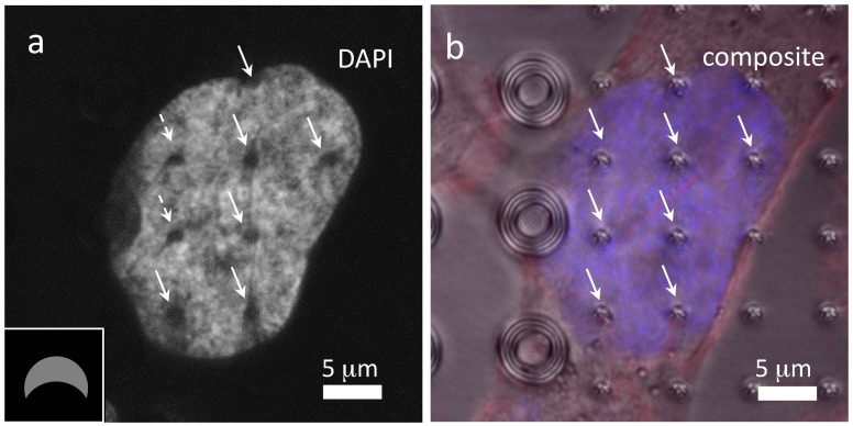
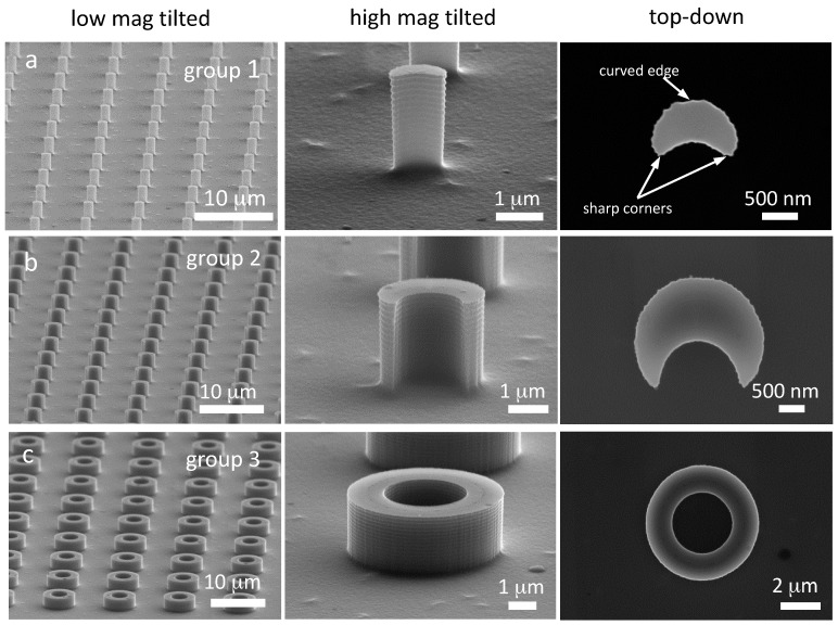
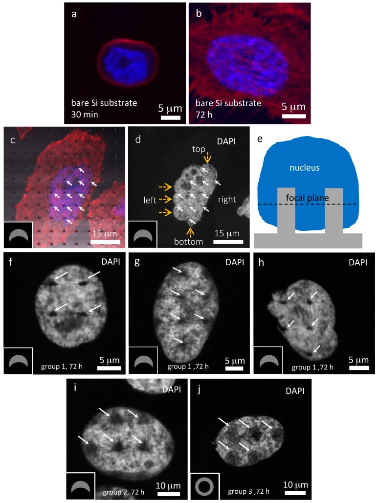
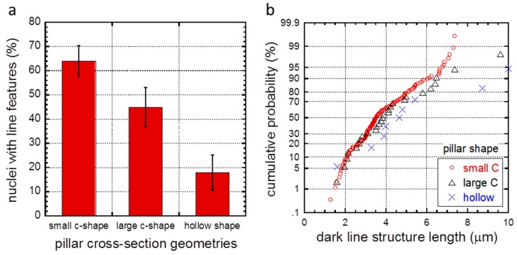
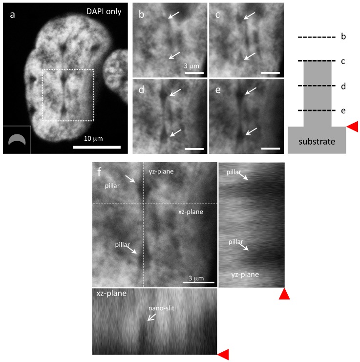
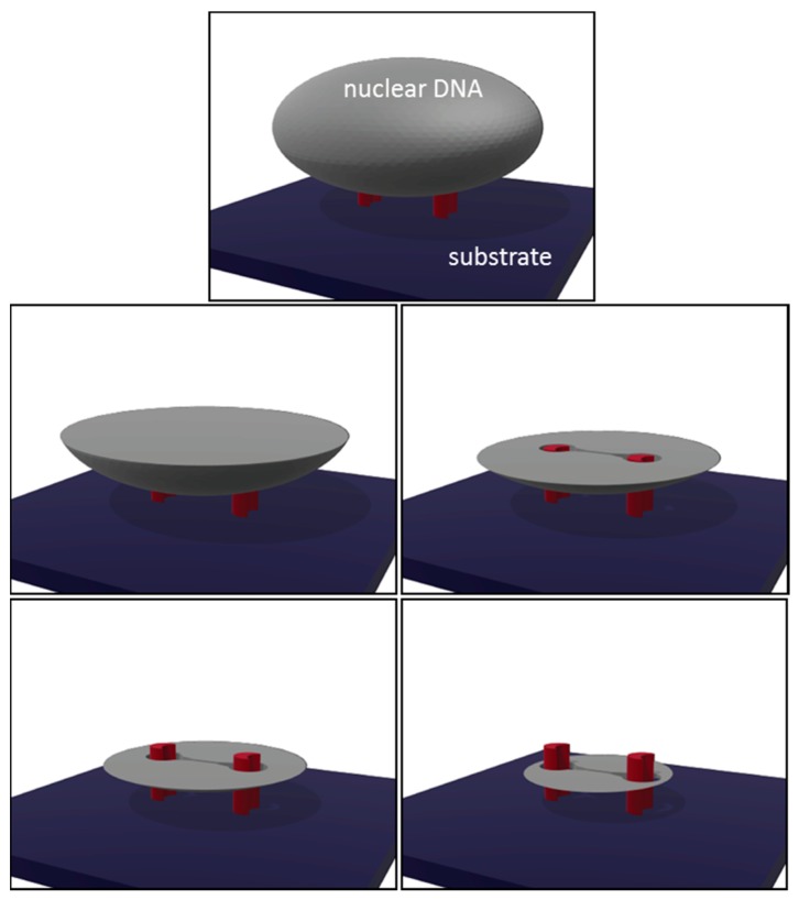
In this study we investigated the contact characteristics of human prostate cancer cells (PC3) on silicon micropillar arrays with complex shapes by using high-resolution confocal fluorescence microscopy techniques. These arrays consist of micropillars that are of various cross-sectional geometries which produce different deformation profiles in adherent cells. Fluorescence micrographs reveal that some DAPI (4',6-diamidino-2-phenylindole)-stained nuclei from cells attached to the pillars develop nanometer scale slits and contain low concentrations of DNA. The lengths of these slits, and their frequency of occurrence, were characterized for various cross-sectional geometries. These DNA-depleted features are only observed in locations below the pillar's top surfaces. Results produced in this study indicate that surface topography can induce unique nanometer scale features in the PC3 cell.
在本研究中,我们使用高分辨率共聚焦荧光显微镜技术,研究了人前列腺癌细胞(PC3)在具有复杂形状的硅微柱阵列上的接触特性。这些阵列由具有各种横截面几何形状的微柱组成,这些微柱在贴壁细胞中产生不同的变形轮廓。荧光显微照片显示,附着在柱子上的细胞中一些经4',6-二脒基-2-苯基吲哚(DAPI)染色的细胞核出现纳米级裂缝,且DNA浓度较低。针对各种横截面几何形状,对这些裂缝的长度及其出现频率进行了表征。这些DNA缺失特征仅在柱子顶面下方的位置观察到。本研究结果表明,表面形貌可在PC3细胞中诱导出独特的纳米级特征。