Korbar Bruno, Olofson Andrea M, Miraflor Allen P, Nicka Catherine M, Suriawinata Matthew A, Torresani Lorenzo, Suriawinata Arief A, Hassanpour Saeed
Department of Biomedical Data Science, Geisel School of Medicine at Dartmouth, One Medical Center Drive, Lebanon, NH 03756, USA.
Department of Computer Science, Dartmouth College, Hanover, NH 03755, USA.
J Pathol Inform. 2017 Jul 25;8:30. doi: 10.4103/jpi.jpi_34_17. eCollection 2017.
Histopathological characterization of colorectal polyps is critical for determining the risk of colorectal cancer and future rates of surveillance for patients. However, this characterization is a challenging task and suffers from significant inter- and intra-observer variability.
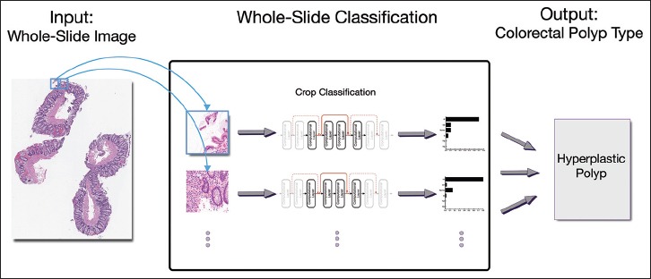
We built an automatic image analysis method that can accurately classify different types of colorectal polyps on whole-slide images to help pathologists with this characterization and diagnosis.
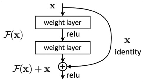
Our method is based on deep-learning techniques, which rely on numerous levels of abstraction for data representation and have shown state-of-the-art results for various image analysis tasks.
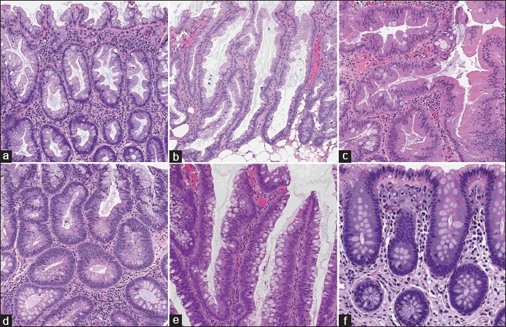
Our method covers five common types of polyps (i.e., hyperplastic, sessile serrated, traditional serrated, tubular, and tubulovillous/villous) that are included in the US Multisociety Task Force guidelines for colorectal cancer risk assessment and surveillance. We developed multiple deep-learning approaches by leveraging a dataset of 2074 crop images, which were annotated by multiple domain expert pathologists as reference standards.
We evaluated our method on an independent test set of 239 whole-slide images and measured standard machine-learning evaluation metrics of accuracy, precision, recall, and F1 score and their 95% confidence intervals.
Our evaluation shows that our method with residual network architecture achieves the best performance for classification of colorectal polyps on whole-slide images (overall accuracy: 93.0%, 95% confidence interval: 89.0%-95.9%).
Our method can reduce the cognitive burden on pathologists and improve their efficacy in histopathological characterization of colorectal polyps and in subsequent risk assessment and follow-up recommendations.
结直肠息肉的组织病理学特征对于确定结直肠癌风险以及患者未来的监测频率至关重要。然而,这种特征描述是一项具有挑战性的任务,并且存在显著的观察者间和观察者内差异。
我们构建了一种自动图像分析方法,该方法可以在全切片图像上准确分类不同类型的结直肠息肉,以帮助病理学家进行这种特征描述和诊断。
我们的方法基于深度学习技术,该技术依赖于多个抽象层次来进行数据表示,并且在各种图像分析任务中都取得了领先的成果。
我们的方法涵盖了美国多学会工作组结直肠癌风险评估和监测指南中包含的五种常见息肉类型(即增生性、无蒂锯齿状、传统锯齿状、管状以及管状绒毛状/绒毛状)。我们利用一个由2074张裁剪图像组成的数据集开发了多种深度学习方法,这些图像由多位领域专家病理学家标注作为参考标准。
我们在一个包含239张全切片图像的独立测试集上评估了我们的方法,并测量了准确性、精确率、召回率和F1分数等标准机器学习评估指标及其95%置信区间。
我们的评估表明,采用残差网络架构的方法在全切片图像上对结直肠息肉进行分类时表现最佳(总体准确率:93.0%,95%置信区间:89.0%-95.9%)。
我们的方法可以减轻病理学家的认知负担,并提高他们在结直肠息肉组织病理学特征描述以及后续风险评估和随访建议方面的效率。