Adenwalla Sherna F, Graham-Brown Matthew P M, Leone Francesca M T, Burton James O, McCann Gerry P
Department of Cardiovascular Sciences, University of Leicester and the NIHR Leicester Cardiovascular Biomedical Research Unit, Glenfield Hospital, Leicester, UK.
John Walls Renal Unit, University Hospitals Leicester NHS Trust, Leicester, UK.
Clin Kidney J. 2017 Aug;10(4):503-515. doi: 10.1093/ckj/sfx028. Epub 2017 May 10.
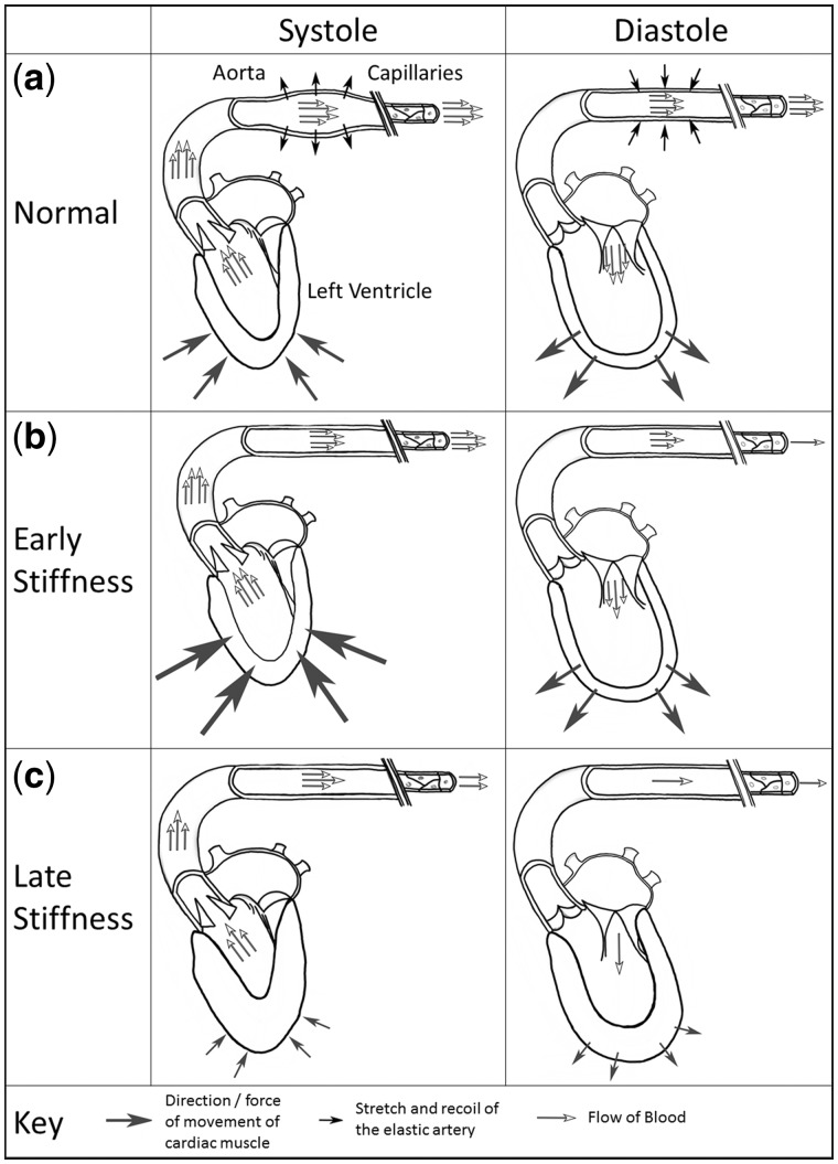
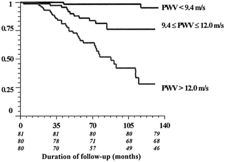
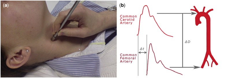
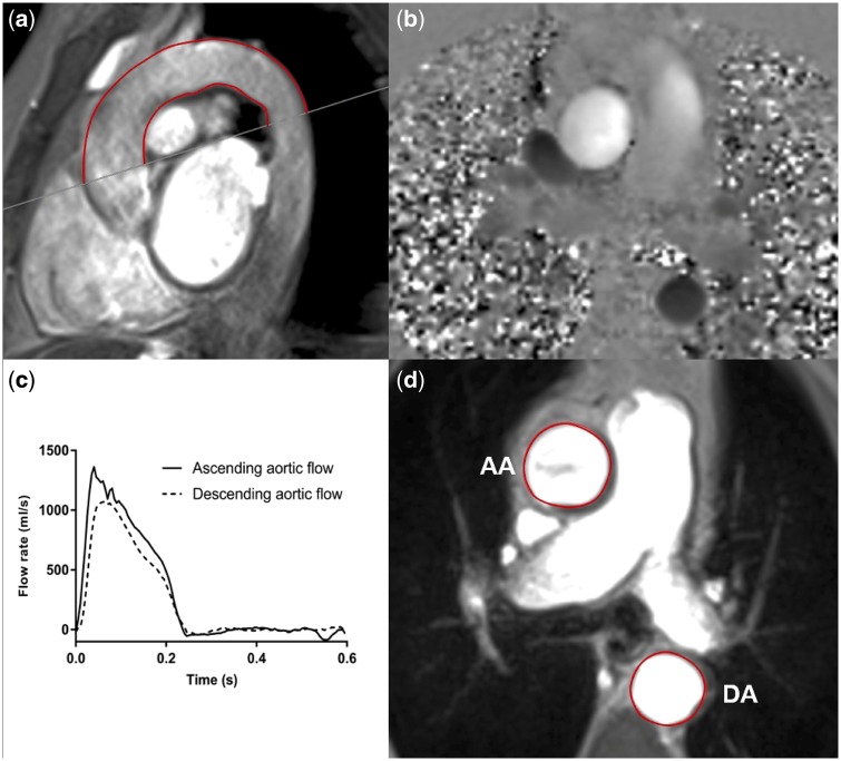
Cardiovascular (CV) disease is the leading cause of death in chronic kidney disease (CKD) and end-stage renal disease (ESRD). A key driver in this pathology is increased aortic stiffness, which is a strong, independent predictor of CV mortality in this population. Aortic stiffening is a potentially modifiable biomarker of CV dysfunction and in risk stratification for patients with CKD and ESRD. Previous work has suggested that therapeutic modification of aortic stiffness may ameliorate CV mortality. Nevertheless, future clinical implementation relies on the ability to accurately and reliably quantify stiffness in renal disease. Pulse wave velocity (PWV) is an indirect measure of stiffness and is the accepted standard for non-invasive assessment of aortic stiffness. It has typically been measured using techniques such as applanation tonometry, which is easy to use but hindered by issues such as the inability to visualize the aorta. Advances in cardiac magnetic resonance imaging now allow direct measurement of stiffness, using aortic distensibility, in addition to PWV. These techniques allow measurement of aortic stiffness locally and are obtainable as part of a comprehensive, multiparametric CV assessment. The evidence cannot yet provide a definitive answer regarding which technique or parameter can be considered superior. This review discusses the advantages and limitations of non-invasive methods that have been used to assess aortic stiffness, the key studies that have assessed aortic stiffness in patients with renal disease and why these tools should be standardized for use in clinical trial work.
心血管(CV)疾病是慢性肾脏病(CKD)和终末期肾病(ESRD)的主要死因。这种病理状态的一个关键驱动因素是主动脉僵硬度增加,这是该人群心血管死亡率的一个强有力的独立预测指标。主动脉硬化是心血管功能障碍的一个潜在可改变的生物标志物,也是CKD和ESRD患者风险分层的指标。先前的研究表明,对主动脉僵硬度进行治疗性改善可能会降低心血管死亡率。然而,未来的临床应用依赖于在肾病中准确可靠地量化僵硬度的能力。脉搏波速度(PWV)是僵硬度的一种间接测量方法,是主动脉僵硬度无创评估的公认标准。它通常使用诸如压平式眼压计等技术进行测量,这种技术易于使用,但存在无法可视化主动脉等问题。心脏磁共振成像的进展现在除了PWV外,还允许使用主动脉扩张性直接测量僵硬度。这些技术可以局部测量主动脉僵硬度,并且可作为全面的多参数心血管评估的一部分获得。关于哪种技术或参数可被认为更优越,现有证据尚未给出明确答案。本综述讨论了用于评估主动脉僵硬度的非侵入性方法的优缺点;评估肾病患者主动脉僵硬度的关键研究;以及为什么这些工具应在临床试验工作中进行标准化使用。