• 文献检索
  • 文档翻译
  • 深度研究
  • 学术资讯
  • Suppr Zotero 插件Zotero 插件
  • 邀请有礼
  • 套餐&价格
  • 历史记录
应用&插件
Suppr Zotero 插件Zotero 插件浏览器插件Mac 客户端Windows 客户端微信小程序
定价
高级版会员购买积分包购买API积分包
服务
文献检索文档翻译深度研究API 文档MCP 服务
关于我们
关于 Suppr公司介绍联系我们用户协议隐私条款
关注我们

Suppr 超能文献

核心技术专利:CN118964589B侵权必究
粤ICP备2023148730 号-1Suppr @ 2026

文献检索

告别复杂PubMed语法,用中文像聊天一样搜索,搜遍4000万医学文献。AI智能推荐,让科研检索更轻松。

立即免费搜索

文件翻译

保留排版,准确专业,支持PDF/Word/PPT等文件格式,支持 12+语言互译。

免费翻译文档

深度研究

AI帮你快速写综述,25分钟生成高质量综述,智能提取关键信息,辅助科研写作。

立即免费体验

一种联合的 3D-SIM/SMLM 方法可以实现以约 4-5nm 的精度对中心体蛋白进行定位。

A combined 3D-SIM/SMLM approach allows centriole proteins to be localized with a precision of ∼4-5 nm.

机构信息

Sir William Dunn School of Pathology, University of Oxford, Oxford, UK.

Micron Oxford Advanced Bioimaging Unit, Department of Biochemistry, University of Oxford, Oxford, UK.

出版信息

Curr Biol. 2017 Oct 9;27(19):R1054-R1055. doi: 10.1016/j.cub.2017.08.009.

DOI:10.1016/j.cub.2017.08.009
PMID:29017036
原文链接:https://pmc.ncbi.nlm.nih.gov/articles/PMC5640508/
Abstract

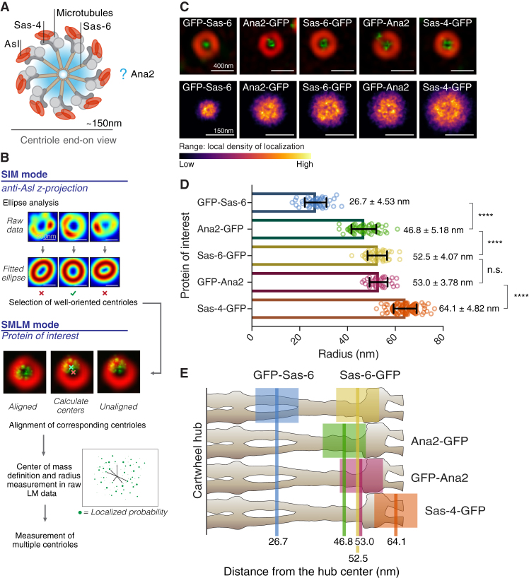
Centrioles are small barrel-shaped structures that form centrosomes and cilia [1]. Centrioles assemble around a central cartwheel comprising the Sas-6 and Ana2/STIL proteins. The amino termini of nine Sas-6 dimers form a central hub of ∼12 nm radius from which nine dimer spokes radiate, placing the Sas-6 carboxyl termini at the outer edge of the ∼60 nm radius cartwheel [2]. Several centriole proteins are distributed in a toroid around the cartwheel, and super-resolution light microscopy studies have measured the average radii of these ∼100-200 nm radius toroids with a 'precision' - or standard deviation (s.d. or 1σ) - of ±∼10-40 nm. The organization of Ana2/STIL within the cartwheel, however, has not been resolvable. Here, we develop methods to calculate the average toroidal radius of centriolar proteins in the ∼20-60 nm range with a s.d. of just ±∼4-5 nm, revealing that the amino and carboxyl termini of Ana2 are located in the outer cartwheel region.

摘要

中心粒是形成中心体和纤毛的小型桶状结构[1]。中心粒围绕由 Sas-6 和 Ana2/STIL 蛋白组成的中央轮辐组装。九个 Sas-6 二聚体的氨基末端形成一个约 12nm 半径的中央轮毂,九个二聚体辐条从这里辐射,将 Sas-6 羧基末端放置在约 60nm 半径轮辐的外边缘[2]。几种中心粒蛋白以轮辐为中心呈环形分布,超分辨率荧光显微镜研究已经测量了这些约 100-200nm 半径轮辐的平均半径,其“精度”或标准偏差(s.d.或 1σ)为±∼10-40nm。然而,轮辐内 Ana2/STIL 的组织尚未得到解决。在这里,我们开发了一种方法,可以计算约 20-60nm 范围内中心粒蛋白的平均环面半径,其标准偏差仅为±∼4-5nm,表明 Ana2 的氨基和羧基末端位于外轮辐区域。

https://cdn.ncbi.nlm.nih.gov/pmc/blobs/d23c/5640508/90c8aba33544/gr1.jpg

相似文献

1
A combined 3D-SIM/SMLM approach allows centriole proteins to be localized with a precision of ∼4-5 nm.一种联合的 3D-SIM/SMLM 方法可以实现以约 4-5nm 的精度对中心体蛋白进行定位。
Curr Biol. 2017 Oct 9;27(19):R1054-R1055. doi: 10.1016/j.cub.2017.08.009.
2
DSas-6 and Ana2 coassemble into tubules to promote centriole duplication and engagement.DSas-6 和 Ana2 共组装形成小管,以促进中心体复制和衔接。
Dev Cell. 2010 Dec 14;19(6):913-9. doi: 10.1016/j.devcel.2010.11.010.
3
The homo-oligomerisation of both Sas-6 and Ana2 is required for efficient centriole assembly in flies.在果蝇中,Sas-6和Ana2的同源寡聚化对于高效的中心粒组装是必需的。
Elife. 2015 May 23;4:e07236. doi: 10.7554/eLife.07236.
4
Drosophila Cep135/Bld10 maintains proper centriole structure but is dispensable for cartwheel formation.果蝇 Cep135/Bld10 维持适当的中心粒结构,但对于轮辐形成是可有可无的。
J Cell Sci. 2012 Dec 1;125(Pt 23):5881-6. doi: 10.1242/jcs.113506. Epub 2012 Sep 12.
5
Cartwheel assembly.侧手翻组件
Philos Trans R Soc Lond B Biol Sci. 2014 Sep 5;369(1650). doi: 10.1098/rstb.2013.0458.
6
SAS-6 is a cartwheel protein that establishes the 9-fold symmetry of the centriole.SAS-6是一种形成中心粒九重对称性的轮状蛋白。
Curr Biol. 2007 Dec 18;17(24):2169-74. doi: 10.1016/j.cub.2007.11.046.
7
The Rise of the Cartwheel: Seeding the Centriole Organelle.车轮的崛起:中心粒细胞器的种子。
Bioessays. 2018 Apr;40(4):e1700241. doi: 10.1002/bies.201700241. Epub 2018 Mar 6.
8
SAS-6 engineering reveals interdependence between cartwheel and microtubules in determining centriole architecture.SAS-6 工程揭示了轮辐蛋白与微管之间在决定中心体结构方面的相互依赖性。
Nat Cell Biol. 2016 Apr;18(4):393-403. doi: 10.1038/ncb3329. Epub 2016 Mar 21.
9
Identification of Chlamydomonas Central Core Centriolar Proteins Reveals a Role for Human WDR90 in Ciliogenesis.鉴定衣藻中心粒核心蛋白揭示了人类 WDR90 在纤毛发生中的作用。
Curr Biol. 2017 Aug 21;27(16):2486-2498.e6. doi: 10.1016/j.cub.2017.07.011. Epub 2017 Aug 3.
10
A molecular mechanism for the procentriole recruitment of Ana2.Ana2向原中心粒募集的分子机制。
J Cell Biol. 2020 Feb 3;219(2). doi: 10.1083/jcb.201905172.

引用本文的文献

1
The A-C linker controls centriole structural integrity and duplication.A-C连接体控制中心粒的结构完整性和复制。
Nat Commun. 2025 Jul 24;16(1):6836. doi: 10.1038/s41467-025-62154-6.
2
Time-series reconstruction of the molecular architecture of human centriole assembly.人类中心体组装的分子结构的时间序列重建。
Cell. 2024 Apr 25;187(9):2158-2174.e19. doi: 10.1016/j.cell.2024.03.025. Epub 2024 Apr 10.
3
Single-molecule imaging in the primary cilium.初级纤毛中单分子成像。

本文引用的文献

1
Asterless licenses daughter centrioles to duplicate for the first time in Drosophila embryos.在果蝇胚胎中,无星蛋白首次许可子中心粒进行复制。
Curr Biol. 2014 Jun 2;24(11):1276-82. doi: 10.1016/j.cub.2014.04.023. Epub 2014 May 15.
2
Structural analysis of the G-box domain of the microcephaly protein CPAP suggests a role in centriole architecture.微脑症蛋白 CPAP 的 G 框结构域的结构分析提示其在中心粒结构中的作用。
Structure. 2013 Nov 5;21(11):2069-77. doi: 10.1016/j.str.2013.08.019. Epub 2013 Sep 26.
3
Crystal structures of the CPAP/STIL complex reveal its role in centriole assembly and human microcephaly.
Methods Cell Biol. 2023;176:59-83. doi: 10.1016/bs.mcb.2023.01.003. Epub 2023 Feb 24.
4
Molecular architecture of the C. elegans centriole.秀丽隐杆线虫中心体的分子结构。
PLoS Biol. 2022 Sep 15;20(9):e3001784. doi: 10.1371/journal.pbio.3001784. eCollection 2022 Sep.
5
Mapping molecular complexes with super-resolution microscopy and single-particle analysis.利用超分辨率显微镜和单颗粒分析技术绘制分子复合物图谱。
Open Biol. 2022 Jul;12(7):220079. doi: 10.1098/rsob.220079. Epub 2022 Jul 27.
6
Centriole distal-end proteins CP110 and Cep97 influence centriole cartwheel growth at the proximal end.中心体远端蛋白 CP110 和 Cep97 影响近端中心体车轮辐状结构的生长。
J Cell Sci. 2022 Jul 15;135(14). doi: 10.1242/jcs.260015. Epub 2022 Jul 20.
7
3D-Structured Illumination Microscopy of Centrosomes in Human Cell Lines.人细胞系中心体的3D结构光照显微镜观察
Bio Protoc. 2022 Mar 20;12(6):e4360. doi: 10.21769/BioProtoc.4360.
8
ENKD1 promotes CP110 removal through competing with CEP97 to initiate ciliogenesis.ENKD1 通过与 CEP97 竞争促进 CP110 的去除来起始纤毛发生。
EMBO Rep. 2022 May 4;23(5):e54090. doi: 10.15252/embr.202154090. Epub 2022 Mar 18.
9
Superresolution characterization of core centriole architecture.核心中心体结构的超分辨率表征。
J Cell Biol. 2021 Apr 5;220(4). doi: 10.1083/jcb.202005103.
10
Sas-4 Colocalizes with the Ciliary Rootlets of the Sensory Organs.Sas-4与感觉器官的纤毛小根共定位。
J Dev Biol. 2021 Jan 5;9(1):1. doi: 10.3390/jdb9010001.
CPAP/STIL复合物的晶体结构揭示了其在中心粒组装和人类小头畸形中的作用。
Elife. 2013 Sep 17;2:e01071. doi: 10.7554/eLife.01071.
4
Native architecture of the centriole proximal region reveals features underlying its 9-fold radial symmetry.中心粒近端区域的固有结构揭示了其 9 重辐射状对称的基础特征。
Curr Biol. 2013 Sep 9;23(17):1620-8. doi: 10.1016/j.cub.2013.06.061. Epub 2013 Aug 8.
5
Subdiffraction-resolution fluorescence microscopy reveals a domain of the centrosome critical for pericentriolar material organization.亚衍射分辨率荧光显微镜揭示了一个对中心体周围物质组织至关重要的中心体域。
Nat Cell Biol. 2012 Nov;14(11):1159-68. doi: 10.1038/ncb2597. Epub 2012 Oct 21.
6
Subdiffraction imaging of centrosomes reveals higher-order organizational features of pericentriolar material.中心体的亚衍射成像揭示了中心粒周围物质的更高阶组织特征。
Nat Cell Biol. 2012 Nov;14(11):1148-58. doi: 10.1038/ncb2591. Epub 2012 Oct 21.
7
Structured illumination of the interface between centriole and peri-centriolar material.中心体和中心粒周围物质界面的结构照明。
Open Biol. 2012 Aug;2(8):120104. doi: 10.1098/rsob.120104.
8
Drosophila Cep135/Bld10 maintains proper centriole structure but is dispensable for cartwheel formation.果蝇 Cep135/Bld10 维持适当的中心粒结构,但对于轮辐形成是可有可无的。
J Cell Sci. 2012 Dec 1;125(Pt 23):5881-6. doi: 10.1242/jcs.113506. Epub 2012 Sep 12.
9
Centrioles, centrosomes, and cilia in health and disease.健康与疾病中的中心粒、中心体和纤毛。
Cell. 2009 Nov 13;139(4):663-78. doi: 10.1016/j.cell.2009.10.036.
10
Imaging intracellular fluorescent proteins at nanometer resolution.以纳米分辨率成像细胞内荧光蛋白。
Science. 2006 Sep 15;313(5793):1642-5. doi: 10.1126/science.1127344. Epub 2006 Aug 10.