• 文献检索
  • 文档翻译
  • 深度研究
  • 学术资讯
  • Suppr Zotero 插件Zotero 插件
  • 邀请有礼
  • 套餐&价格
  • 历史记录
应用&插件
Suppr Zotero 插件Zotero 插件浏览器插件Mac 客户端Windows 客户端微信小程序
定价
高级版会员购买积分包购买API积分包
服务
文献检索文档翻译深度研究API 文档MCP 服务
关于我们
关于 Suppr公司介绍联系我们用户协议隐私条款
关注我们

Suppr 超能文献

核心技术专利:CN118964589B侵权必究
粤ICP备2023148730 号-1Suppr @ 2026

文献检索

告别复杂PubMed语法,用中文像聊天一样搜索,搜遍4000万医学文献。AI智能推荐,让科研检索更轻松。

立即免费搜索

文件翻译

保留排版,准确专业,支持PDF/Word/PPT等文件格式,支持 12+语言互译。

免费翻译文档

深度研究

AI帮你快速写综述,25分钟生成高质量综述,智能提取关键信息,辅助科研写作。

立即免费体验

核心中心体结构的超分辨率表征。

Superresolution characterization of core centriole architecture.

机构信息

State Key Laboratory of Agrobiotechnology, College of Biological Sciences, China Agricultural University, Beijing, China.

Tsinghua-Peking Joint Center for Life Sciences and Max Planck Partner Group, School of Life Sciences, Tsinghua University, Beijing, China.

出版信息

J Cell Biol. 2021 Apr 5;220(4). doi: 10.1083/jcb.202005103.

DOI:10.1083/jcb.202005103
PMID:33533934
原文链接:https://pmc.ncbi.nlm.nih.gov/articles/PMC7863704/
Abstract

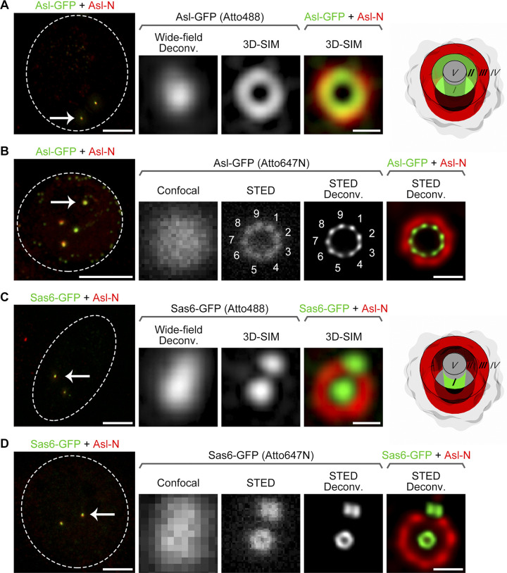
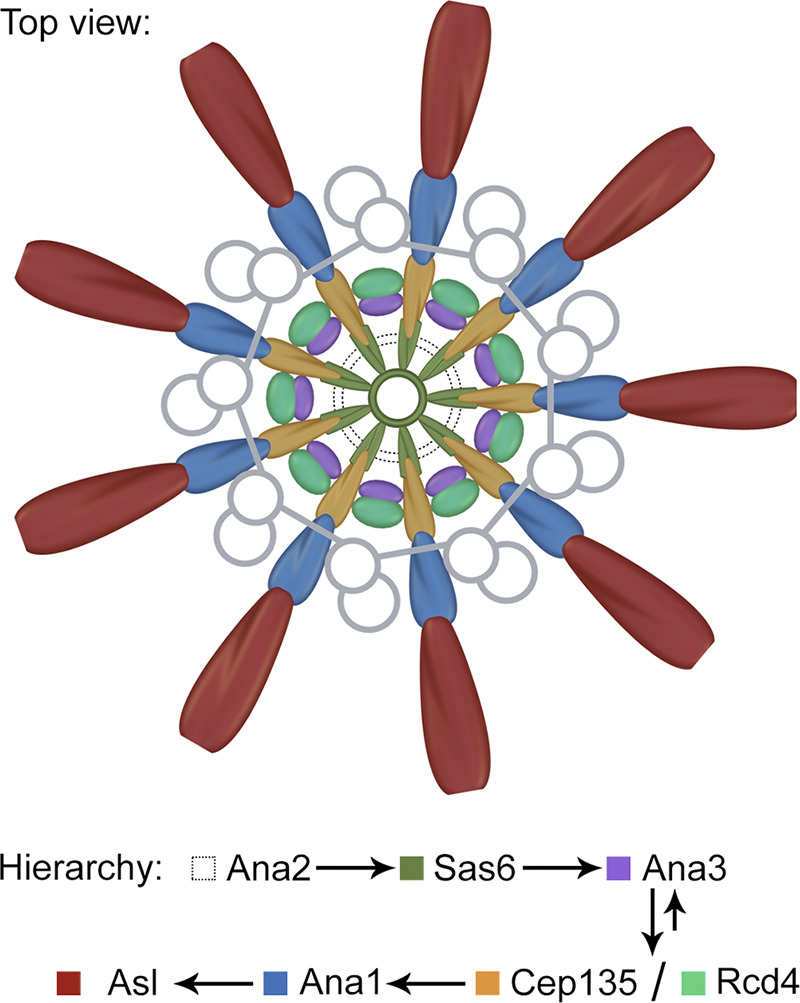
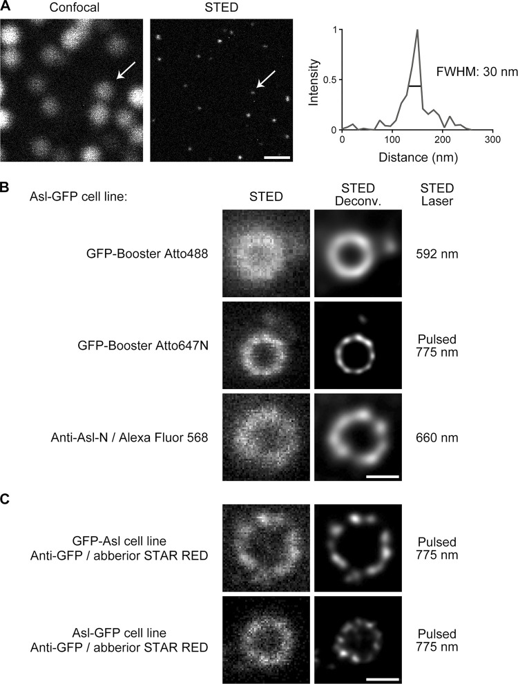
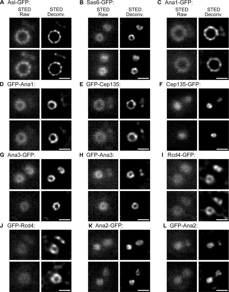
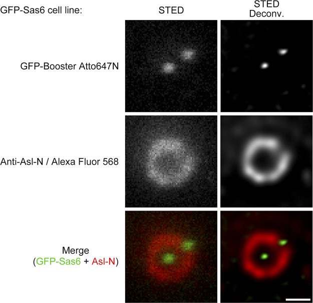
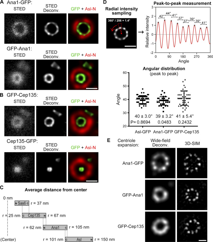
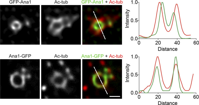
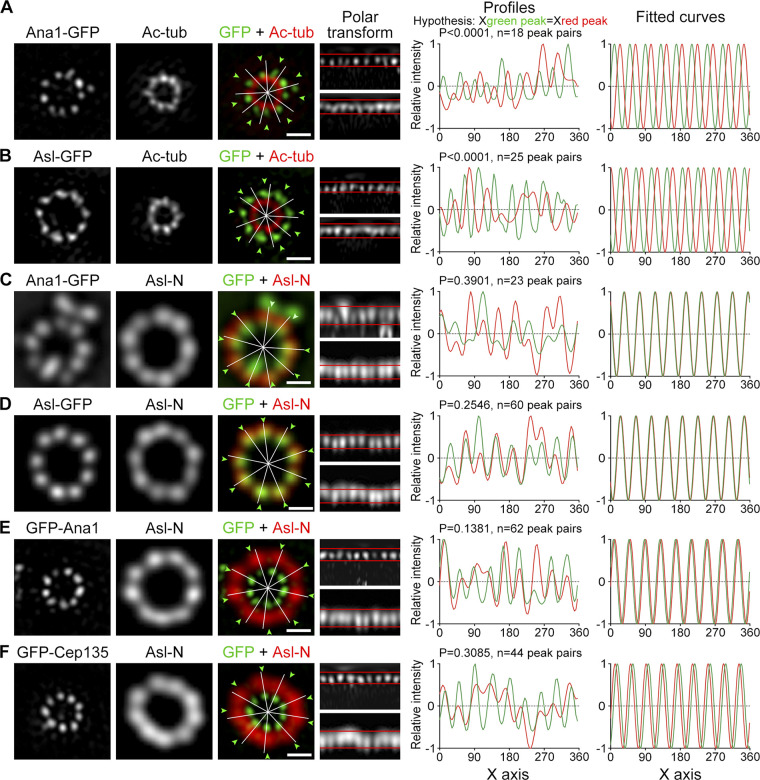
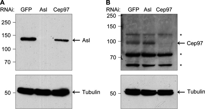
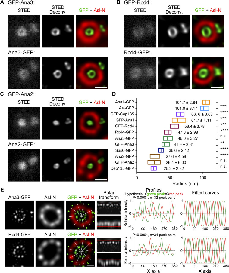
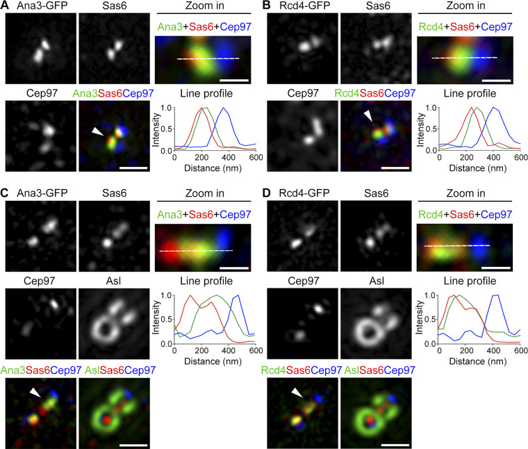
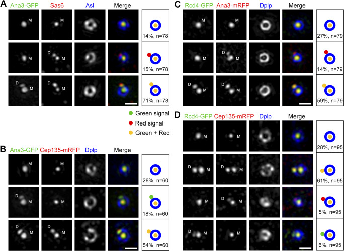
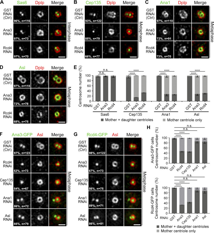
The centrosome is the main microtubule-organizing center in animal cells. It comprises of two centrioles and the surrounding pericentriolar material. Protein organization at the outer layer of the centriole and outward has been studied extensively; however, an overall picture of the protein architecture at the centriole core has been missing. Here we report a direct view of Drosophila centriolar proteins at ∼50-nm resolution. This reveals a Sas6 ring at the C-terminus, where it overlaps with the C-terminus of Cep135. The ninefold symmetrical pattern of Cep135 is further conveyed through Ana1-Asterless axes that extend past the microtubule wall from between the blades. Ana3 and Rcd4, whose termini are close to Cep135, are arranged in ninefold symmetry that does not match the above axes. During centriole biogenesis, Ana3 and Rcd4 are sequentially loaded on the newly formed centriole and are required for centriole-to-centrosome conversion through recruiting the Cep135-Ana1-Asterless complex. Together, our results provide a spatiotemporal map of the centriole core and implications of how the structure might be built.

摘要

中心体是动物细胞中主要的微管组织中心。它由两个中心粒和周围的中心粒周围物质组成。中心粒外层和向外的蛋白质组织已经得到了广泛的研究;然而,中心粒核心的蛋白质结构的全貌仍然缺失。在这里,我们报告了约 50nm 分辨率的果蝇中心粒蛋白的直接观察结果。这揭示了 Sas6 环在 C 末端,在那里它与 Cep135 的 C 末端重叠。Cep135 的九倍对称模式进一步通过 Ana1-Asterless 轴传递,该轴从叶片之间延伸到微管壁之外。靠近 Cep135 的 Ana3 和 Rcd4 以九倍对称排列,与上述轴不匹配。在中心体发生过程中,Ana3 和 Rcd4 依次加载到新形成的中心粒上,并通过招募 Cep135-Ana1-Asterless 复合物,对中心体到中心体的转化是必需的。总之,我们的结果提供了中心粒核心的时空图谱,并暗示了结构可能是如何构建的。

https://cdn.ncbi.nlm.nih.gov/pmc/blobs/c942/7863704/80b67651cc3b/JCB_202005103_Fig8.jpg
https://cdn.ncbi.nlm.nih.gov/pmc/blobs/c942/7863704/08b6a25dc5ca/JCB_202005103_Fig1.jpg
https://cdn.ncbi.nlm.nih.gov/pmc/blobs/c942/7863704/8e018bddf94c/JCB_202005103_FigS1.jpg
https://cdn.ncbi.nlm.nih.gov/pmc/blobs/c942/7863704/78c7a27bed2d/JCB_202005103_FigS2.jpg
https://cdn.ncbi.nlm.nih.gov/pmc/blobs/c942/7863704/21e897c2d3bf/JCB_202005103_FigS3.jpg
https://cdn.ncbi.nlm.nih.gov/pmc/blobs/c942/7863704/868658aef413/JCB_202005103_Fig2.jpg
https://cdn.ncbi.nlm.nih.gov/pmc/blobs/c942/7863704/3dd572c4c8c3/JCB_202005103_FigS4.jpg
https://cdn.ncbi.nlm.nih.gov/pmc/blobs/c942/7863704/eeefc287eb0f/JCB_202005103_Fig3.jpg
https://cdn.ncbi.nlm.nih.gov/pmc/blobs/c942/7863704/65e7447fa519/JCB_202005103_FigS5.jpg
https://cdn.ncbi.nlm.nih.gov/pmc/blobs/c942/7863704/849de6740d62/JCB_202005103_Fig4.jpg
https://cdn.ncbi.nlm.nih.gov/pmc/blobs/c942/7863704/99b61751601a/JCB_202005103_Fig5.jpg
https://cdn.ncbi.nlm.nih.gov/pmc/blobs/c942/7863704/28d2a69e9ebc/JCB_202005103_Fig6.jpg
https://cdn.ncbi.nlm.nih.gov/pmc/blobs/c942/7863704/3b8f0e9ba0df/JCB_202005103_Fig7.jpg
https://cdn.ncbi.nlm.nih.gov/pmc/blobs/c942/7863704/80b67651cc3b/JCB_202005103_Fig8.jpg

相似文献

1
Superresolution characterization of core centriole architecture.核心中心体结构的超分辨率表征。
J Cell Biol. 2021 Apr 5;220(4). doi: 10.1083/jcb.202005103.
2
9-fold symmetry is not essential for centriole elongation and formation of new centriole-like structures.九倍对称对于中心体的伸长和新的中心体样结构的形成不是必需的。
Nat Commun. 2024 May 25;15(1):4467. doi: 10.1038/s41467-024-48831-y.
3
Drosophila Ana1 is required for centrosome assembly and centriole elongation.果蝇Ana1是中心体组装和中心粒延长所必需的。
J Cell Sci. 2016 Jul 1;129(13):2514-25. doi: 10.1242/jcs.186460. Epub 2016 May 20.
4
Drosophila Cep135/Bld10 maintains proper centriole structure but is dispensable for cartwheel formation.果蝇 Cep135/Bld10 维持适当的中心粒结构,但对于轮辐形成是可有可无的。
J Cell Sci. 2012 Dec 1;125(Pt 23):5881-6. doi: 10.1242/jcs.113506. Epub 2012 Sep 12.
5
Conserved molecular interactions in centriole-to-centrosome conversion.中心粒到中心体转化过程中保守的分子相互作用。
Nat Cell Biol. 2016 Jan;18(1):87-99. doi: 10.1038/ncb3274. Epub 2015 Nov 23.
6
Asterless is a scaffold for the onset of centriole assembly.无星状体蛋白是中心体组装起始的支架。
Nature. 2010 Oct 7;467(7316):714-8. doi: 10.1038/nature09445. Epub 2010 Sep 19.
7
Structured illumination of the interface between centriole and peri-centriolar material.中心体和中心粒周围物质界面的结构照明。
Open Biol. 2012 Aug;2(8):120104. doi: 10.1098/rsob.120104.
8
DSAS-6 organizes a tube-like centriole precursor, and its absence suggests modularity in centriole assembly.DSAS-6 组织形成一种管状中心粒前体,其缺失表明中心粒组装具有模块化特征。
Curr Biol. 2007 Sep 4;17(17):1465-72. doi: 10.1016/j.cub.2007.07.034. Epub 2007 Aug 9.
9
Tissue specific requirement of Drosophila Rcd4 for centriole duplication and ciliogenesis.果蝇 Rcd4 对中心体复制和纤毛发生的组织特异性需求。
J Cell Biol. 2020 Sep 7;219(8). doi: 10.1083/jcb.201912154.
10
The origin of the second centriole in the zygote of Drosophila melanogaster.黑腹果蝇合子中第二个中心粒的起源。
Genetics. 2014 May;197(1):199-205. doi: 10.1534/genetics.113.160523. Epub 2014 Feb 13.

引用本文的文献

1
The structure of basal body inner junctions from Tetrahymena revealed by electron cryo-tomography.冷冻电子断层扫描揭示的嗜热四膜虫基体内部连接结构。
EMBO J. 2025 Apr;44(7):1975-2001. doi: 10.1038/s44318-025-00392-6. Epub 2025 Feb 24.
2
Interactions of N- and C-terminal parts of Ana1 permitting centriole duplication but not elongation.Ana1的N端和C端部分相互作用,允许中心粒复制但不允许其延长。
Open Biol. 2025 Feb;15(2):240325. doi: 10.1098/rsob.240325. Epub 2025 Feb 5.
3
Interactions of N- and C-terminal parts of Ana1 permitting centriole duplication but not elongation.

本文引用的文献

1
Architecture of the centriole cartwheel-containing region revealed by cryo-electron tomography.中心粒轮辐结构域的冷冻电镜断层成像图
EMBO J. 2020 Nov 16;39(22):e106246. doi: 10.15252/embj.2020106246. Epub 2020 Sep 20.
2
Tissue specific requirement of Drosophila Rcd4 for centriole duplication and ciliogenesis.果蝇 Rcd4 对中心体复制和纤毛发生的组织特异性需求。
J Cell Biol. 2020 Sep 7;219(8). doi: 10.1083/jcb.201912154.
3
High-resolution characterization of centriole distal appendage morphology and dynamics by correlative STORM and electron microscopy.
Ana1的N端和C端部分的相互作用允许中心粒复制但不允许其延长。
bioRxiv. 2024 Oct 31:2024.10.28.620588. doi: 10.1101/2024.10.28.620588.
4
The Structure of Cilium Inner Junctions Revealed by Electron Cryo-tomography.冷冻电子断层扫描揭示纤毛内部连接的结构
bioRxiv. 2024 Sep 9:2024.09.09.612100. doi: 10.1101/2024.09.09.612100.
5
CP110 and CEP135 Localize Near the Proximal Centriolar Remnants of Mice Spermatozoa.CP110和CEP135定位于小鼠精子近端中心粒残余物附近。
MicroPubl Biol. 2024 Jan 29;2024. doi: 10.17912/micropub.biology.001083. eCollection 2024.
6
Ana1/CEP295 is an essential player in the centrosome maintenance program regulated by Polo kinase and the PCM.CEP295 蛋白是中心体维持的关键蛋白,受 Polo 激酶和 PCM 调控。
EMBO Rep. 2024 Jan;25(1):102-127. doi: 10.1038/s44319-023-00020-6. Epub 2024 Jan 10.
7
A simple Turing reaction-diffusion model explains how PLK4 breaks symmetry during centriole duplication and assembly.一个简单的图灵反应扩散模型解释了 PLK4 如何在中心体复制和组装过程中打破对称。
PLoS Biol. 2023 Nov 20;21(11):e3002391. doi: 10.1371/journal.pbio.3002391. eCollection 2023 Nov.
8
Towards understanding centriole elimination.为了理解中心体的消除。
Open Biol. 2023 Nov;13(11):230222. doi: 10.1098/rsob.230222. Epub 2023 Nov 15.
9
The Endothelial Centrosome: Specific Features and Functional Significance for Endothelial Cell Activity and Barrier Maintenance.内皮中心体:内皮细胞活性和屏障维持的特定特征和功能意义。
Int J Mol Sci. 2023 Oct 20;24(20):15392. doi: 10.3390/ijms242015392.
10
Immunolabel-First-Expand-Later Expansion Microscopy Approach Using Stable STED Dyes.基于稳定 STED 染料的免疫标记-先扩张后标记显微镜方法。
Methods Mol Biol. 2024;2725:89-101. doi: 10.1007/978-1-0716-3507-0_5.
通过共聚焦 STORM 和电子显微镜对中心粒远端附属物形态和动力学进行高分辨率表征。
Nat Commun. 2019 Mar 1;10(1):993. doi: 10.1038/s41467-018-08216-4.
4
Mechanism and Regulation of Centriole and Cilium Biogenesis.中心体和纤毛发生的机制与调控。
Annu Rev Biochem. 2019 Jun 20;88:691-724. doi: 10.1146/annurev-biochem-013118-111153. Epub 2019 Jan 11.
5
Imaging cellular ultrastructures using expansion microscopy (U-ExM).使用扩展显微镜(U-ExM)对细胞超微结构进行成像。
Nat Methods. 2019 Jan;16(1):71-74. doi: 10.1038/s41592-018-0238-1. Epub 2018 Dec 17.
6
PPP1R35 ensures centriole homeostasis by promoting centriole-to-centrosome conversion.PPP1R35 通过促进中心体转化来确保中心体的稳态。
Mol Biol Cell. 2018 Nov 15;29(23):2801-2808. doi: 10.1091/mbc.E18-08-0525. Epub 2018 Sep 19.
7
PPP1R35 is a novel centrosomal protein that regulates centriole length in concert with the microcephaly protein RTTN.PPP1R35 是一种新型中心体蛋白,与小头畸形蛋白 RTTN 一起调节中心粒长度。
Elife. 2018 Aug 31;7:e37846. doi: 10.7554/eLife.37846.
8
Insights into centriole geometry revealed by cryotomography of doublet and triplet centrioles.通过对二联体和三联体中心粒的冷冻断层成像技术揭示中心粒的几何结构。
Elife. 2018 Aug 6;7:e36851. doi: 10.7554/eLife.36851.
9
Super-resolution architecture of mammalian centriole distal appendages reveals distinct blade and matrix functional components.哺乳动物中心粒远端附属物的超分辨率结构揭示了不同的叶片和基质功能组件。
Nat Commun. 2018 May 22;9(1):2023. doi: 10.1038/s41467-018-04469-1.
10
STED nanoscopy of the centrosome linker reveals a CEP68-organized, periodic rootletin network anchored to a C-Nap1 ring at centrioles.基于 STED 的中心体连接蛋白超分辨成像揭示了一个由 CEP68 组织的、有规律的根丝蛋白网络,该网络锚定于中心粒的 C-Nap1 环上。
Proc Natl Acad Sci U S A. 2018 Mar 6;115(10):E2246-E2253. doi: 10.1073/pnas.1716840115. Epub 2018 Feb 20.