• 文献检索
  • 文档翻译
  • 深度研究
  • 学术资讯
  • Suppr Zotero 插件Zotero 插件
  • 邀请有礼
  • 套餐&价格
  • 历史记录
应用&插件
Suppr Zotero 插件Zotero 插件浏览器插件Mac 客户端Windows 客户端微信小程序
定价
高级版会员购买积分包购买API积分包
服务
文献检索文档翻译深度研究API 文档MCP 服务
关于我们
关于 Suppr公司介绍联系我们用户协议隐私条款
关注我们

Suppr 超能文献

核心技术专利:CN118964589B侵权必究
粤ICP备2023148730 号-1Suppr @ 2026

文献检索

告别复杂PubMed语法,用中文像聊天一样搜索,搜遍4000万医学文献。AI智能推荐,让科研检索更轻松。

立即免费搜索

文件翻译

保留排版,准确专业,支持PDF/Word/PPT等文件格式,支持 12+语言互译。

免费翻译文档

深度研究

AI帮你快速写综述,25分钟生成高质量综述,智能提取关键信息,辅助科研写作。

立即免费体验

羟氯喹毒性导致的黄斑周围指状交叉区缺失在近红外反射成像上表现为亚临床靶心病变。

Perifoveal interdigitation zone loss in hydroxychloroquine toxicity leads to subclinical bull's eye lesion appearance on near-infrared reflectance imaging.

作者信息

Chew Avenell L, Sampson Danuta M, Chelva Enid, Khan Jane C, Chen Fred K

机构信息

Centre for Ophthalmology and Visual Science (Incorporating Lions Eye Institute), The University of Western Australia, Crawley, WA, 6009, Australia.

Department of Medical Technology and Physics, Sir Charles Gairdner Hospital, Nedlands, WA, 6009, Australia.

出版信息

Doc Ophthalmol. 2018 Feb;136(1):57-68. doi: 10.1007/s10633-017-9615-9. Epub 2017 Nov 9.

DOI:10.1007/s10633-017-9615-9
PMID:29124422
原文链接:https://pmc.ncbi.nlm.nih.gov/articles/PMC5811575/
Abstract

PURPOSE

To characterize the ultrastructural and functional correlates of hydroxychloroquine (HCQ)-induced subclinical bull's eye lesion seen on near-infrared reflectance (NIR) imaging.

METHODS

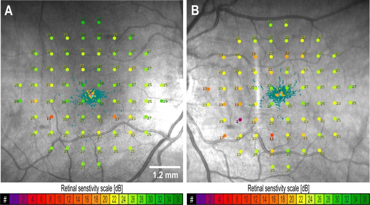
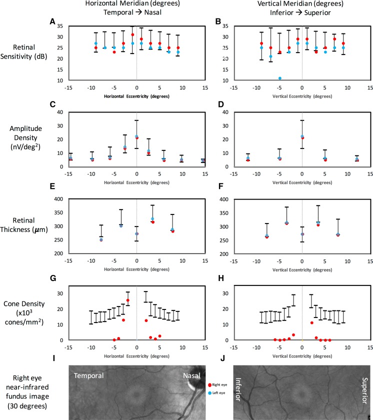
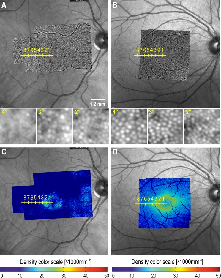
An asymptomatic 54-year-old male taking HCQ presented with paracentral ring-like scotoma, abnormal multifocal electroretinography (mfERG) and preserved ellipsoid zone on optical coherence tomography (OCT). Dense raster OCT was performed to create en face reflectivity maps of the interdigitation zone. Macular Integrity Assessment (MAIA) microperimetry and mfERG findings were compared with NIR imaging, en face OCT, retinal thickness profiles and wave-guiding cone density maps derived from flood-illumination adaptive optics (AO) retinal photography.

RESULTS

The bull's eye lesion is an oval annular zone of increased reflectivity on NIR with an outer diameter of 1450 µm. This region corresponds exactly to an area of preserved interdigitation zone reflectivity in en face OCT images and of normal cone density on AO imaging. Immediately surrounding the bull's eye lesion is an annular zone (3°-12° eccentricity) of depressed retinal sensitivity on MAIA and reduced amplitude density on mfERG. Wave-guiding cone density at 2° temporal was 25,400 per mm. This declined rapidly to 12,900 and 1200 per mm at 3° and 4°.

CONCLUSION

Multimodal imaging illustrated pathology in the area surrounding the NIR bull's eye, characterized by reduced reflectance, wave-guiding cone density and retinal function. Further studies are required to investigate whether the bull's eye on NIR imaging and en face OCT is prominent or consistent enough for diagnostic use.

摘要

目的

描述在近红外反射(NIR)成像中观察到的羟氯喹(HCQ)诱导的亚临床靶心病变的超微结构和功能相关性。

方法

一名无症状的54岁男性服用HCQ后出现旁中心环状暗点、异常多焦视网膜电图(mfERG)以及光学相干断层扫描(OCT)上的椭圆体带保存。进行密集光栅OCT以创建指状交叉区的正面反射率图。将黄斑完整性评估(MAIA)微视野检查和mfERG结果与NIR成像、正面OCT、视网膜厚度剖面图以及源自泛光照明自适应光学(AO)视网膜摄影的波导锥密度图进行比较。

结果

靶心病变是NIR上反射率增加的椭圆形环形区域,外径为1450 µm。该区域与正面OCT图像中保存的指状交叉区反射率区域以及AO成像上的正常视锥密度区域完全对应。紧挨着靶心病变的是一个环形区域(偏心度3° - 12°),在MAIA上视网膜敏感度降低,在mfERG上振幅密度降低。颞侧2°处的波导锥密度为每毫米25,400个。在3°和4°处迅速降至每毫米12,900个和1200个。

结论

多模态成像显示了NIR靶心周围区域的病理情况,其特征为反射率、波导锥密度和视网膜功能降低。需要进一步研究来调查NIR成像和正面OCT上的靶心是否突出或一致到足以用于诊断。

https://cdn.ncbi.nlm.nih.gov/pmc/blobs/fd8a/5811575/ad0ea854186b/10633_2017_9615_Fig5_HTML.jpg
https://cdn.ncbi.nlm.nih.gov/pmc/blobs/fd8a/5811575/79a87121beb8/10633_2017_9615_Fig1_HTML.jpg
https://cdn.ncbi.nlm.nih.gov/pmc/blobs/fd8a/5811575/77f5ff3816d7/10633_2017_9615_Fig2_HTML.jpg
https://cdn.ncbi.nlm.nih.gov/pmc/blobs/fd8a/5811575/c85e735aa640/10633_2017_9615_Fig3_HTML.jpg
https://cdn.ncbi.nlm.nih.gov/pmc/blobs/fd8a/5811575/a24157727599/10633_2017_9615_Fig4_HTML.jpg
https://cdn.ncbi.nlm.nih.gov/pmc/blobs/fd8a/5811575/ad0ea854186b/10633_2017_9615_Fig5_HTML.jpg

相似文献

1
Perifoveal interdigitation zone loss in hydroxychloroquine toxicity leads to subclinical bull's eye lesion appearance on near-infrared reflectance imaging.羟氯喹毒性导致的黄斑周围指状交叉区缺失在近红外反射成像上表现为亚临床靶心病变。
Doc Ophthalmol. 2018 Feb;136(1):57-68. doi: 10.1007/s10633-017-9615-9. Epub 2017 Nov 9.
2
A possible early sign of hydroxychloroquine macular toxicity.羟氯喹黄斑毒性的一种可能早期迹象。
Doc Ophthalmol. 2016 Feb;132(1):75-81. doi: 10.1007/s10633-015-9521-y. Epub 2016 Jan 20.
3
Effect of disease stage on progression of hydroxychloroquine retinopathy.疾病阶段对羟氯喹视网膜病变进展的影响。
JAMA Ophthalmol. 2014 Sep;132(9):1105-12. doi: 10.1001/jamaophthalmol.2014.1099.
4
Early hydroxychloroquine retinopathy: optical coherence tomography abnormalities preceding Humphrey visual field defects.早期羟氯喹性视网膜病变:Humphrey 视野缺损前的光学相干断层扫描异常。
Br J Ophthalmol. 2019 Nov;103(11):1600-1604. doi: 10.1136/bjophthalmol-2018-313350. Epub 2019 Feb 28.
5
Disparity between visual fields and optical coherence tomography in hydroxychloroquine retinopathy.视野与光学相干断层扫描在羟氯喹相关性视网膜病变中的差异。
Ophthalmology. 2014 Jun;121(6):1257-62. doi: 10.1016/j.ophtha.2013.12.002. Epub 2014 Jan 16.
6
Assessment of parafoveal cone density in patients taking hydroxychloroquine in the absence of clinically documented retinal toxicity.在无临床记录视网膜毒性的情况下,对服用羟氯喹的患者进行旁中心凹视锥细胞密度评估。
Acta Ophthalmol. 2015 Nov;93(7):e534-40. doi: 10.1111/aos.12728. Epub 2015 May 20.
7
Case Report: Hydroxychloroquine Retinopathy.病例报告:羟氯喹视网膜病变
Optom Vis Sci. 2018 Jun;95(6):545-549. doi: 10.1097/OPX.0000000000001226.
8
Retinal toxicity of high-dose hydroxychloroquine in patients with chronic graft-versus-host disease.高剂量羟氯喹对慢性移植物抗宿主病患者的视网膜毒性
Can J Ophthalmol. 2015 Dec;50(6):442-50. doi: 10.1016/j.jcjo.2015.08.003.
9
Multimodal imaging in a severe case of hydroxychloroquine toxicity.羟氯喹毒性严重病例中的多模态成像
Ophthalmic Surg Lasers Imaging Retina. 2015 Mar;46(3):377-9. doi: 10.3928/23258160-20150323-14.
10
Enhanced Visualization of Subtle Outer Retinal Pathology by En Face Optical Coherence Tomography and Correlation with Multi-Modal Imaging.通过表面光学相干断层扫描增强对视网膜外层细微病变的可视化及其与多模态成像的相关性
PLoS One. 2016 Dec 13;11(12):e0168275. doi: 10.1371/journal.pone.0168275. eCollection 2016.

引用本文的文献

1
Censoring the Floor Effect in Long-Term Stargardt Disease Microperimetry Data Produces a Faster Rate of Decline.对长期Stargardt病微视野检查数据中的下限效应进行审查会导致更快的下降速度。
Ophthalmol Sci. 2024 Jul 20;4(6):100581. doi: 10.1016/j.xops.2024.100581. eCollection 2024 Nov-Dec.
2
Pearls and Pitfalls of Adaptive Optics Ophthalmoscopy in Inherited Retinal Diseases.遗传性视网膜疾病中自适应光学检眼镜检查的要点与陷阱
Diagnostics (Basel). 2023 Jul 19;13(14):2413. doi: 10.3390/diagnostics13142413.
3
Review of Retinal Imaging Modalities for Hydroxychloroquine Retinopathy.

本文引用的文献

1
Semi-automated identification of cones in the human retina using circle Hough transform.使用圆形霍夫变换对人眼视网膜中的视锥细胞进行半自动识别。
Biomed Opt Express. 2015 Nov 3;6(12):4676-93. doi: 10.1364/BOE.6.004676. eCollection 2015 Dec 1.
2
The Negative Cone Mosaic: A New Manifestation of the Optical Stiles-Crawford Effect in Normal Eyes.负锥镶嵌:正常眼睛中光学斯泰尔斯-克劳福德效应的一种新表现形式。
Invest Ophthalmol Vis Sci. 2015 Nov;56(12):7043-50. doi: 10.1167/iovs.15-17022.
3
Assessment of Different Sampling Methods for Measuring and Representing Macular Cone Density Using Flood-Illuminated Adaptive Optics.
羟氯喹视网膜病变的视网膜成像模式综述
Diagnostics (Basel). 2023 May 16;13(10):1752. doi: 10.3390/diagnostics13101752.
4
Bull's Eye Maculopathy in Near-Infrared Reflectance as An Early Sign of Hydroxychloroquine Toxicity.近红外反射中的靶心样黄斑病变作为羟氯喹毒性的早期迹象
Diagnostics (Basel). 2023 Jan 26;13(3):445. doi: 10.3390/diagnostics13030445.
5
Edge of Scotoma Sensitivity as a Microperimetry Clinical Trial End Point in Retinopathy.视网膜病变中作为微视野临床试验终点的暗点边缘敏感度
Transl Vis Sci Technol. 2020 Sep 9;9(10):9. doi: 10.1167/tvst.9.10.9. eCollection 2020 Sep.
6
Interpreting MAIA Microperimetry Using Age- and Retinal Loci-Specific Reference Thresholds.使用年龄和视网膜位点特异性参考阈值解读MAIA微视野计检查结果
Transl Vis Sci Technol. 2020 Jun 18;9(7):19. doi: 10.1167/tvst.9.7.19. eCollection 2020 Jun.
7
Hydroxychloroquine-Induced Toxic Myopathy Causing Diaphragmatic Weakness and Lung Collapse Requiring Prolonged Mechanical Ventilation.羟氯喹诱导的毒性肌病导致膈肌无力和肺塌陷,需要长时间机械通气。
J Investig Med High Impact Case Rep. 2020 Jan-Dec;8:2324709620950113. doi: 10.1177/2324709620950113.
使用泛光照明自适应光学技术评估测量和表征黄斑视锥细胞密度的不同采样方法
Invest Ophthalmol Vis Sci. 2015 Sep;56(10):5751-63. doi: 10.1167/iovs.15-16954.
4
RETICULAR PSEUDODRUSEN ON INFRARED IMAGING ARE TOPOGRAPHICALLY DISTINCT FROM SUBRETINAL DRUSENOID DEPOSITS ON EN FACE OPTICAL COHERENCE TOMOGRAPHY.红外成像上的网状假性玻璃膜疣在地形学上与正面光学相干断层扫描上的视网膜下类玻璃膜疣沉积物不同。
Retina. 2015 Dec;35(12):2593-603. doi: 10.1097/IAE.0000000000000666.
5
Near-infrared reflectance bull's eye maculopathy as an early indication of hydroxychloroquine toxicity.近红外反射性靶心性黄斑病变作为羟氯喹毒性的早期指征
Clin Ophthalmol. 2015 Mar 19;9:521-5. doi: 10.2147/OPTH.S76963. eCollection 2015.
6
Subjective and objective screening tests for hydroxychloroquine toxicity.羟氯喹毒性的主观和客观筛查试验
Ophthalmology. 2015 Feb;122(2):356-66. doi: 10.1016/j.ophtha.2014.07.056. Epub 2014 Oct 14.
7
Meaning of visualizing retinal cone mosaic on adaptive optics images.在自适应光学图像上可视化视网膜锥细胞镶嵌的意义。
Am J Ophthalmol. 2015 Jan;159(1):118-23.e1. doi: 10.1016/j.ajo.2014.09.043. Epub 2014 Oct 2.
8
Variations in the cone packing density with eccentricity in emmetropes.正视眼中视锥细胞堆积密度随偏心率的变化。
Eye (Lond). 2014 Dec;28(12):1488-93. doi: 10.1038/eye.2014.229. Epub 2014 Oct 3.
9
In vivo imaging of human cone photoreceptor inner segments.人眼视锥细胞内节的活体成像。
Invest Ophthalmol Vis Sci. 2014 Jun 6;55(7):4244-51. doi: 10.1167/iovs.14-14542.
10
Disparity between visual fields and optical coherence tomography in hydroxychloroquine retinopathy.视野与光学相干断层扫描在羟氯喹相关性视网膜病变中的差异。
Ophthalmology. 2014 Jun;121(6):1257-62. doi: 10.1016/j.ophtha.2013.12.002. Epub 2014 Jan 16.