Ghensi Paolo, Bressan Eriberto, Gardin Chiara, Ferroni Letizia, Soldini Maria Costanza, Mandelli Federico, Soldini Claudio, Zavan Barbara
Department of Neurosciences, Dental School, University of Padova, Via Giustiniani 2, 35100 Padova, Italy.
Centre for Integrative Biology (CIBIO), University of Trento, 38122 Trento, Italy.
Materials (Basel). 2017 Nov 17;10(11):1321. doi: 10.3390/ma10111321.
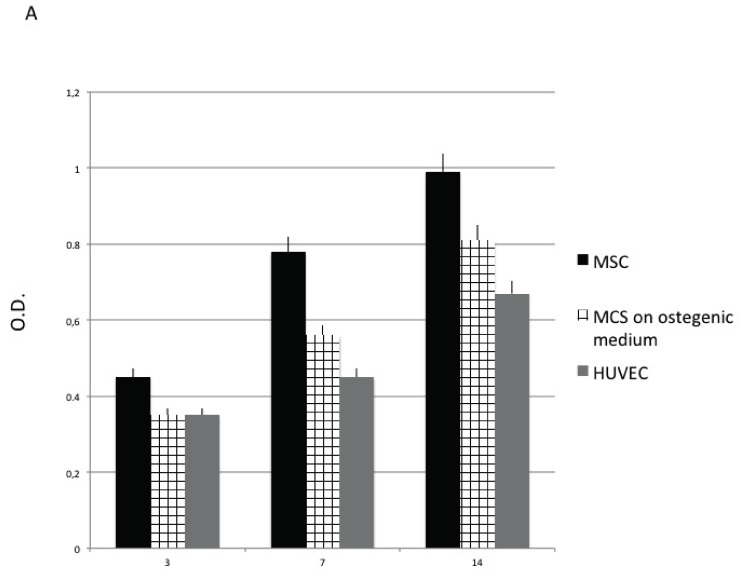
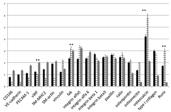
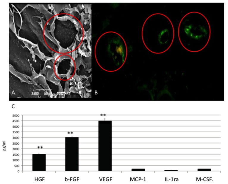
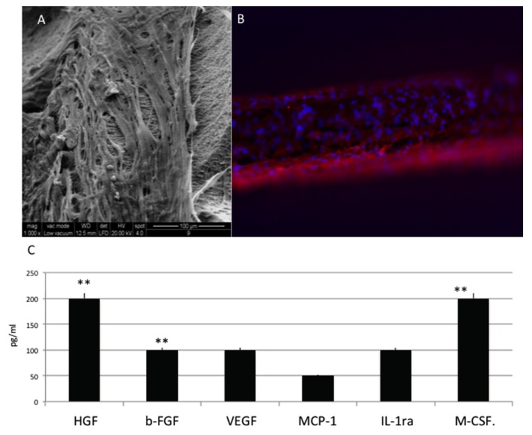
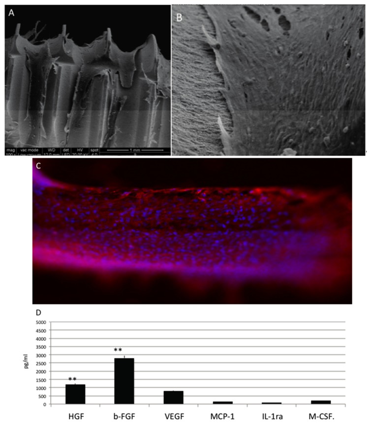
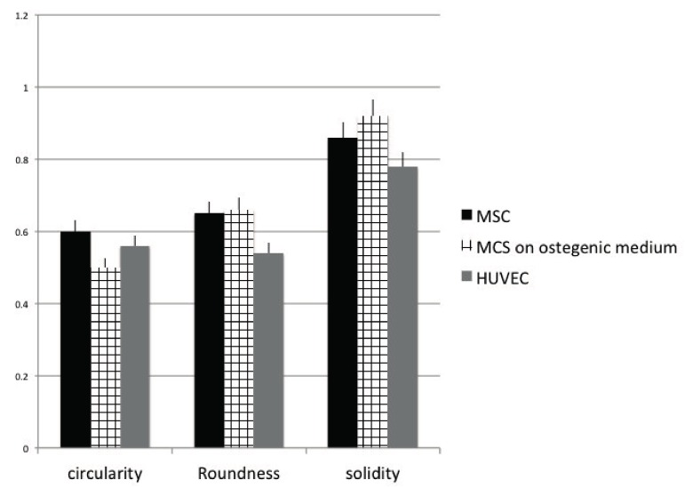
Osteogenesis process displays a fundamental role during dental implant osteointegration. In the present work, we studied the influence of Osteon Growth Induction (OGI) surface properties on the angiogenic and osteogenic behaviors of Mesenchymal Stem cells (MSC). MSC derived from dental pulp and HUVEC (Human Umbilical Vein Endothelial Cells) were grown in on OGI titanium surfaces, and cell proliferation and DNA synthesis were evaluated by MTT [3-(4,5-dimethylthiazol-2yl)-2,5-diphenyltetrazolium bromide] test and DNA quantification. Gene expression has been performed in order to evaluate the presence of mRNA related to endothelial and osteogenesis markers. Moreover, morphological and biochemical analyses of osteogenesis commitments has been performed. On OGI surfaces, MSC and HUVEC are able to proliferate. Gene expression profiler confirms that MSC on OGI surfaces are able to express endothelial and osteogenic markers, and that these expression are higher compared the expression on control surfaces. In conclusion On OGI surfaces proliferation, expression and morphological analyses of angiogenesis-associated markers in MSC are promoted. This process induces an increasing on their osteogenesis commitment.
骨生成过程在牙种植体骨整合中发挥着重要作用。在本研究中,我们研究了骨生长诱导(OGI)表面特性对间充质干细胞(MSC)血管生成和成骨行为的影响。将来自牙髓的MSC和人脐静脉内皮细胞(HUVEC)培养在OGI钛表面,通过MTT [3-(4,5-二甲基噻唑-2)-2,5-二苯基四氮唑溴盐] 试验和DNA定量评估细胞增殖和DNA合成。进行基因表达以评估与内皮和成骨标志物相关的mRNA的存在。此外,还进行了骨生成承诺的形态学和生化分析。在OGI表面上,MSC和HUVEC能够增殖。基因表达谱证实,OGI表面上的MSC能够表达内皮和成骨标志物,并且这些表达比对照表面上的表达更高。总之,在OGI表面上,MSC中与血管生成相关标志物的增殖、表达和形态学分析得到促进。这一过程促使它们的骨生成承诺增加。