• 文献检索
  • 文档翻译
  • 深度研究
  • 学术资讯
  • Suppr Zotero 插件Zotero 插件
  • 邀请有礼
  • 套餐&价格
  • 历史记录
应用&插件
Suppr Zotero 插件Zotero 插件浏览器插件Mac 客户端Windows 客户端微信小程序
定价
高级版会员购买积分包购买API积分包
服务
文献检索文档翻译深度研究API 文档MCP 服务
关于我们
关于 Suppr公司介绍联系我们用户协议隐私条款
关注我们

Suppr 超能文献

核心技术专利:CN118964589B侵权必究
粤ICP备2023148730 号-1Suppr @ 2026

文献检索

告别复杂PubMed语法,用中文像聊天一样搜索,搜遍4000万医学文献。AI智能推荐,让科研检索更轻松。

立即免费搜索

文件翻译

保留排版,准确专业,支持PDF/Word/PPT等文件格式,支持 12+语言互译。

免费翻译文档

深度研究

AI帮你快速写综述,25分钟生成高质量综述,智能提取关键信息,辅助科研写作。

立即免费体验

纳米颗粒表面涂层对细胞毒性和线粒体摄取的影响。

Effect of Nanoparticle Surface Coating on Cell Toxicity and Mitochondria Uptake.

作者信息

Zheng Hong, Mortensen Luke J, Ravichandran Supriya, Bentley Karen, DeLouise Lisa A

出版信息

J Biomed Nanotechnol. 2017 Feb;13(2):155-66. doi: 10.1166/jbn.2017.2337.

DOI:10.1166/jbn.2017.2337
PMID:29377103
原文链接:https://pmc.ncbi.nlm.nih.gov/articles/PMC6800125/
Abstract

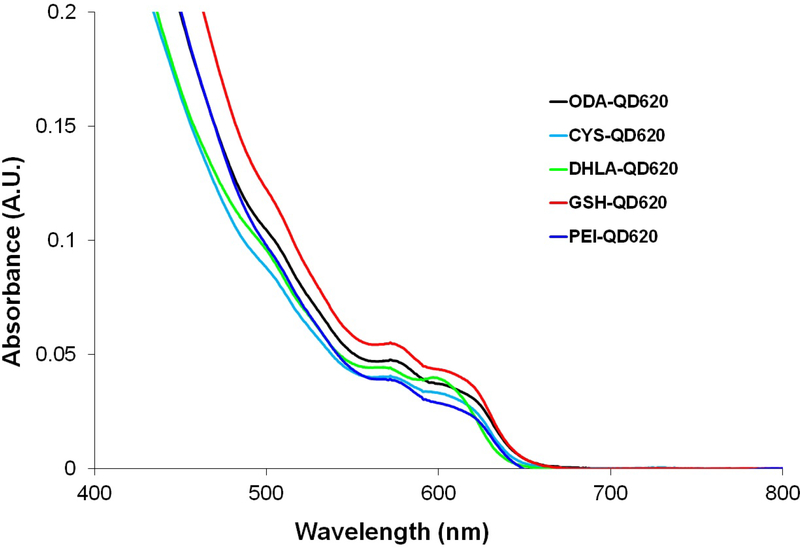
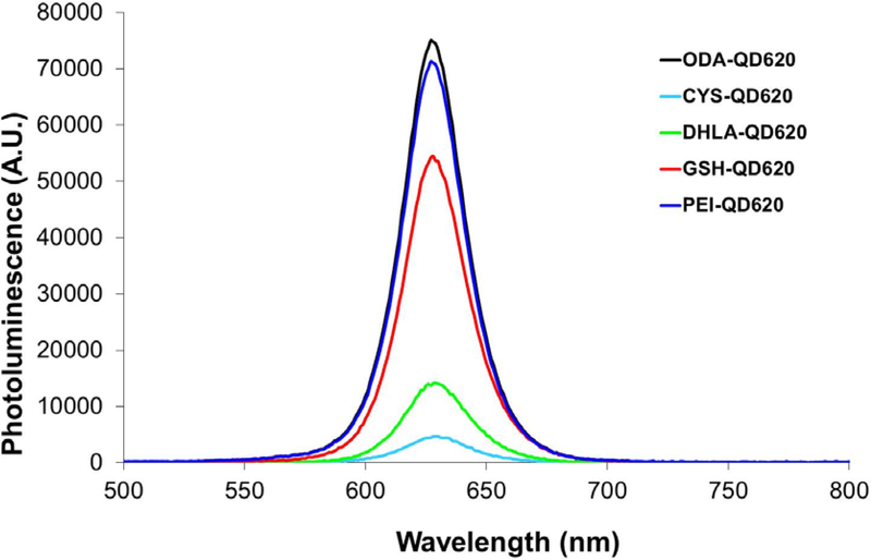
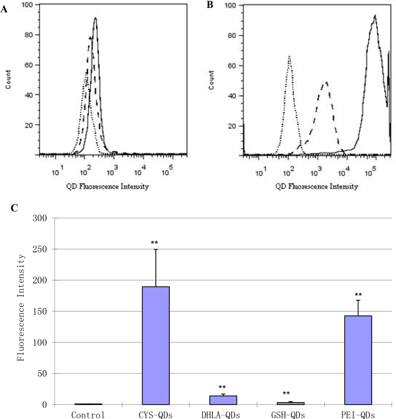
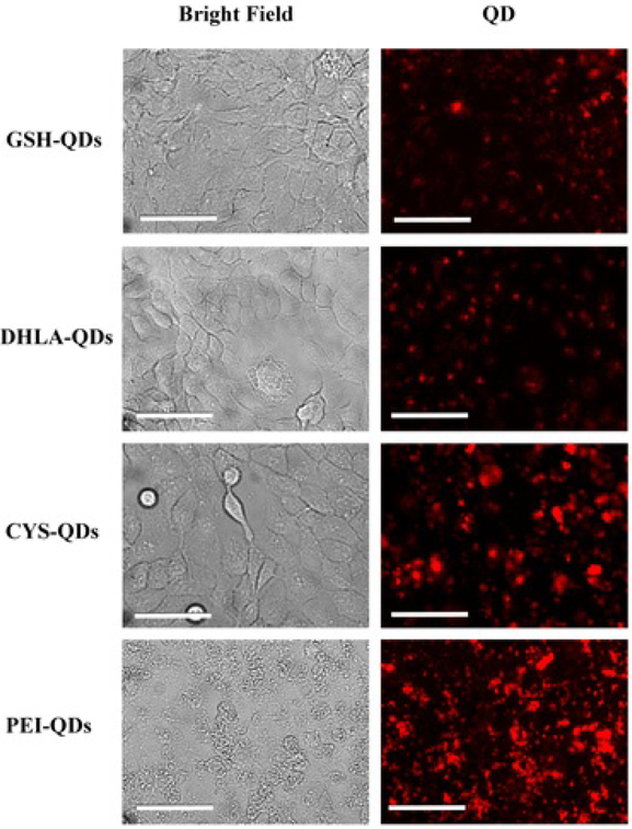
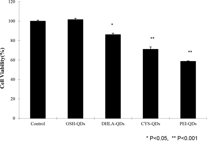
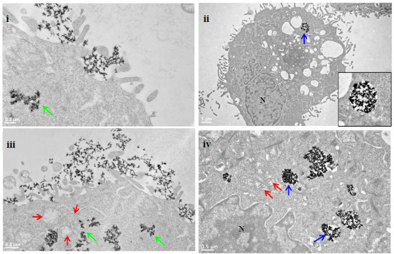
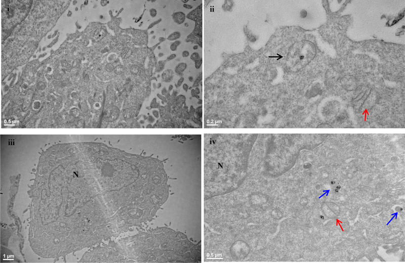
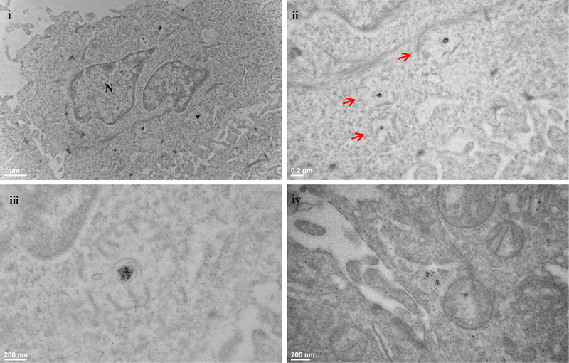
We report on the effect of surface charge and the ligand coating composition of CdSe/ZnS core/shell quantum dot (QD) nanoparticles on human keratinocyte toxicity using fluorescent microscopy, flow cytometry, transmission electron microscopy. Two commonly reported positive charged (cysteamine, polyethylenimine) and two negative charged (glutathione, dihydrolipoic acid) ligands were studied. The QDs were fully characterized by UV-vis absorption spectroscopy, fluorescence emission spectroscopy, dynamic light scattering and zeta potential. Differences in surface coatings and charges were evaluated against cellular uptake, ROS generation, cytotoxicity, and mitochondrial targeting. Results show that the negative charged QDs coated with GSH exhibit excellent water solubility, high quantum yield and low cytotoxicity. Ligand composition is more important in ROS generation than surface charge whereas surface charge is an important driver of cytotoxicity. Most importantly we observe the selective accumulation of glutathione coated QDs in vesicles in the mitochondria matrix. This observation suggests a new strategy for developing mitochondria-targeted nanomaterials for drug/gene delivery.

摘要

我们使用荧光显微镜、流式细胞术、透射电子显微镜,报告了CdSe/ZnS核壳量子点(QD)纳米颗粒的表面电荷和配体涂层组成对人角质形成细胞毒性的影响。研究了两种常见的带正电荷的配体(半胱胺、聚乙烯亚胺)和两种带负电荷的配体(谷胱甘肽、二氢硫辛酸)。通过紫外可见吸收光谱、荧光发射光谱、动态光散射和zeta电位对量子点进行了全面表征。针对细胞摄取、活性氧生成、细胞毒性和线粒体靶向性,评估了表面涂层和电荷的差异。结果表明,包覆谷胱甘肽的带负电荷的量子点具有优异的水溶性、高量子产率和低细胞毒性。配体组成在活性氧生成中比表面电荷更重要,而表面电荷是细胞毒性的重要驱动因素。最重要的是,我们观察到谷胱甘肽包覆的量子点选择性地积累在线粒体基质的囊泡中。这一观察结果为开发用于药物/基因递送的线粒体靶向纳米材料提出了一种新策略。

https://cdn.ncbi.nlm.nih.gov/pmc/blobs/b151/6800125/2897084689e6/nihms-1053945-f0016.jpg
https://cdn.ncbi.nlm.nih.gov/pmc/blobs/b151/6800125/bdc5f1012aef/nihms-1053945-f0006.jpg
https://cdn.ncbi.nlm.nih.gov/pmc/blobs/b151/6800125/e111f6f26be7/nihms-1053945-f0007.jpg
https://cdn.ncbi.nlm.nih.gov/pmc/blobs/b151/6800125/6e2428294f4d/nihms-1053945-f0008.jpg
https://cdn.ncbi.nlm.nih.gov/pmc/blobs/b151/6800125/f883ca7d54ab/nihms-1053945-f0009.jpg
https://cdn.ncbi.nlm.nih.gov/pmc/blobs/b151/6800125/34ce2786a040/nihms-1053945-f0010.jpg
https://cdn.ncbi.nlm.nih.gov/pmc/blobs/b151/6800125/c44dbddddf04/nihms-1053945-f0011.jpg
https://cdn.ncbi.nlm.nih.gov/pmc/blobs/b151/6800125/6112f8c863f1/nihms-1053945-f0012.jpg
https://cdn.ncbi.nlm.nih.gov/pmc/blobs/b151/6800125/0158c8027c45/nihms-1053945-f0013.jpg
https://cdn.ncbi.nlm.nih.gov/pmc/blobs/b151/6800125/f7e916be72e7/nihms-1053945-f0014.jpg
https://cdn.ncbi.nlm.nih.gov/pmc/blobs/b151/6800125/b43cc3f0746d/nihms-1053945-f0015.jpg
https://cdn.ncbi.nlm.nih.gov/pmc/blobs/b151/6800125/2897084689e6/nihms-1053945-f0016.jpg

相似文献

1
Effect of Nanoparticle Surface Coating on Cell Toxicity and Mitochondria Uptake.纳米颗粒表面涂层对细胞毒性和线粒体摄取的影响。
J Biomed Nanotechnol. 2017 Feb;13(2):155-66. doi: 10.1166/jbn.2017.2337.
2
Thiol antioxidant-functionalized CdSe/ZnS quantum dots: synthesis, characterization, cytotoxicity.巯基抗氧化功能化 CdSe/ZnS 量子点:合成、表征、细胞毒性。
J Biomed Nanotechnol. 2013 Mar;9(3):382-92. doi: 10.1166/jbn.2013.1561.
3
Cellular uptake, elimination and toxicity of CdSe/ZnS quantum dots in HepG2 cells.CdSe/ZnS 量子点在 HepG2 细胞中的细胞摄取、消除和毒性。
Biomaterials. 2013 Dec;34(37):9545-58. doi: 10.1016/j.biomaterials.2013.08.038. Epub 2013 Sep 5.
4
The role of surface chemistry in determining in vivo biodistribution and toxicity of CdSe/ZnS core-shell quantum dots.表面化学在决定 CdSe/ZnS 核壳量子点体内分布和毒性中的作用。
Biomaterials. 2013 Nov;34(34):8741-55. doi: 10.1016/j.biomaterials.2013.07.087. Epub 2013 Aug 9.
5
Cytotoxicity of quantum dots used for in vitro cellular labeling: role of QD surface ligand, delivery modality, cell type, and direct comparison to organic fluorophores.用于体外细胞标记的量子点的细胞毒性:QD 表面配体、递送方式、细胞类型的作用,以及与有机荧光染料的直接比较。
Bioconjug Chem. 2013 Sep 18;24(9):1570-83. doi: 10.1021/bc4001917. Epub 2013 Aug 16.
6
In vivo assessment of CdSe-ZnS quantum dots: coating dependent bioaccumulation and genotoxicity.体内评估 CdSe-ZnS 量子点:涂层依赖性生物蓄积和遗传毒性。
Nanoscale. 2012 Oct 21;4(20):6401-7. doi: 10.1039/c2nr31826a.
7
CdSe/ZnS quantum dots exhibited nephrotoxicity through mediating oxidative damage and inflammatory response.硒化镉/硫化锌量子点通过介导氧化损伤和炎症反应表现出肾毒性。
Aging (Albany NY). 2020 Nov 16;13(8):12194-12206. doi: 10.18632/aging.103774.
8
The role of intracellular trafficking of CdSe/ZnS QDs on their consequent toxicity profile.硒化镉/硫化锌量子点的细胞内运输在其后续毒性特征方面的作用。
J Nanobiotechnology. 2017 Jun 15;15(1):45. doi: 10.1186/s12951-017-0279-0.
9
Evaluation of quantum dot cytotoxicity: interpretation of nanoparticle concentrations versus intracellular nanoparticle numbers.量子点细胞毒性评估:纳米颗粒浓度与细胞内纳米颗粒数量的解读
Nanotoxicology. 2016 Nov;10(9):1318-28. doi: 10.1080/17435390.2016.1210691. Epub 2016 Jul 28.
10
Cell type-dependent changes in CdSe/ZnS quantum dot uptake and toxic endpoints.CdSe/ZnS量子点摄取及毒性终点的细胞类型依赖性变化。
Toxicol Sci. 2015 Apr;144(2):246-58. doi: 10.1093/toxsci/kfv002. Epub 2015 Jan 19.

引用本文的文献

1
Surface Charge Affects the Intracellular Fate and Clearance Dynamics of CdSe/ZnS Quantum Dots in Macrophages.表面电荷影响巨噬细胞中CdSe/ZnS量子点的细胞内命运和清除动力学。
Nanomaterials (Basel). 2025 Aug 3;15(15):1189. doi: 10.3390/nano15151189.
2
Recent Breakthroughs in Using Quantum Dots for Cancer Imaging and Drug Delivery Purposes.量子点在癌症成像与药物递送方面的近期突破
Nanomaterials (Basel). 2023 Sep 15;13(18):2566. doi: 10.3390/nano13182566.
3
SiO Fibers of Two Lengths and Their Effect on Cellular Responses of Macrophage-like Cells.

本文引用的文献

1
Glutathione and mitochondria.谷胱甘肽与线粒体。
Front Pharmacol. 2014 Jul 1;5:151. doi: 10.3389/fphar.2014.00151. eCollection 2014.
2
Regulation of intracellular signaling and function by caveolin.小窝蛋白对细胞内信号传导和功能的调节
FASEB J. 2014 Sep;28(9):3823-31. doi: 10.1096/fj.14-252320. Epub 2014 May 22.
3
Polymer-coated nanoparticles interacting with proteins and cells: focusing on the sign of the net charge.聚合物包覆的纳米颗粒与蛋白质和细胞的相互作用:聚焦于净电荷的符号。
两种长度的 SiO 纤维及其对巨噬细胞样细胞细胞反应的影响。
Molecules. 2022 Jul 12;27(14):4456. doi: 10.3390/molecules27144456.
4
One-Step Aqueous Synthesis of Anionic and Cationic AgInS Quantum Dots and Their Utility in Improving the Efficacy of ALA-Based Photodynamic Therapy.一步水相合成阴离子和阳离子 AgInS 量子点及其在提高基于 ALA 的光动力疗法疗效中的应用。
Inorg Chem. 2022 Feb 14;61(6):2846-2863. doi: 10.1021/acs.inorgchem.1c03298. Epub 2022 Feb 1.
5
Insights into Theranostic Properties of Titanium Dioxide for Nanomedicine.二氧化钛在纳米医学中的诊疗特性洞察
Nanomicro Lett. 2020 Jan 14;12(1):22. doi: 10.1007/s40820-019-0362-1.
6
In vivo Comparison of the Biodistribution and Toxicity of InP/ZnS Quantum Dots with Different Surface Modifications.体内比较不同表面修饰的 InP/ZnS 量子点的生物分布和毒性。
Int J Nanomedicine. 2020 Mar 20;15:1951-1965. doi: 10.2147/IJN.S241332. eCollection 2020.
7
Cardiotoxicity of Intravenously Administered CdSe/ZnS Quantum Dots in BALB/c Mice.静脉注射CdSe/ZnS量子点对BALB/c小鼠的心脏毒性
Front Pharmacol. 2019 Oct 8;10:1179. doi: 10.3389/fphar.2019.01179. eCollection 2019.
8
Recent Advances in Mitochondria-Targeted Gene Delivery.线粒体靶向基因传递的最新进展。
Molecules. 2018 Sep 11;23(9):2316. doi: 10.3390/molecules23092316.
9
Cytotoxicity of InP/ZnS Quantum Dots With Different Surface Functional Groups Toward Two Lung-Derived Cell Lines.具有不同表面官能团的InP/ZnS量子点对两种肺源细胞系的细胞毒性
Front Pharmacol. 2018 Jul 13;9:763. doi: 10.3389/fphar.2018.00763. eCollection 2018.
ACS Nano. 2013 Apr 23;7(4):3253-63. doi: 10.1021/nn3059295. Epub 2013 Apr 8.
4
Apoptosis-induced mitochondrial dysfunction causes cytoplasmic lipid droplet formation.细胞凋亡诱导的线粒体功能障碍导致细胞质脂滴形成。
Cell Death Differ. 2012 Sep;19(9):1561-70. doi: 10.1038/cdd.2012.34. Epub 2012 Mar 30.
5
A vesicular transport pathway shuttles cargo from mitochondria to lysosomes.囊泡运输途径将货物从线粒体运输到溶酶体。
Curr Biol. 2012 Jan 24;22(2):135-41. doi: 10.1016/j.cub.2011.11.057. Epub 2012 Jan 5.
6
Cellular uptake, intracellular trafficking, and cytotoxicity of nanomaterials.纳米材料的细胞摄取、细胞内转运和细胞毒性。
Small. 2011 May 23;7(10):1322-37. doi: 10.1002/smll.201100001. Epub 2011 Apr 26.
7
Quantum dots, lighting up the research and development of nanomedicine.量子点,点亮纳米医学研发之路。
Nanomedicine. 2011 Aug;7(4):385-402. doi: 10.1016/j.nano.2010.12.006. Epub 2011 Jan 5.
8
Endo- and exocytosis of zwitterionic quantum dot nanoparticles by live HeLa cells.活 HeLa 细胞内两性离子量子点纳米颗粒的内吞作用和胞吐作用。
ACS Nano. 2010 Nov 23;4(11):6787-97. doi: 10.1021/nn101277w. Epub 2010 Oct 28.
9
Nanoscale surface modifications of medically relevant metals: state-of-the art and perspectives.医学相关金属的纳米级表面改性:现状与展望。
Nanoscale. 2011 Feb;3(2):335-53. doi: 10.1039/c0nr00485e. Epub 2010 Oct 26.
10
Gold nanoparticles and quantum dots for bioimaging.金纳米粒子和量子点用于生物成像。
Microsc Res Tech. 2011 Jul;74(7):592-604. doi: 10.1002/jemt.20928. Epub 2010 Sep 9.