Müller Elena K, Gräfe Christine, Wiekhorst Frank, Bergemann Christian, Weidner Andreas, Dutz Silvio, Clement Joachim H
Department Hematology and Oncology, Jena University Hospital, Am Klinikum 1, D-07747 Jena, Germany.
Physikalisch-Technische Bundesanstalt Berlin, Abbestr. 2-12, D-10587 Berlin, Germany.
Nanomaterials (Basel). 2018 Feb 14;8(2):108. doi: 10.3390/nano8020108.
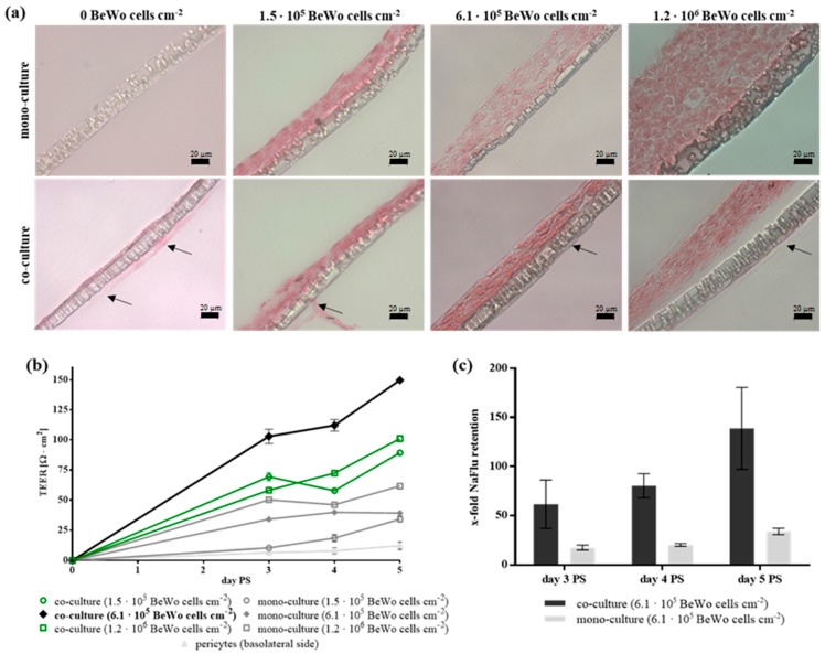
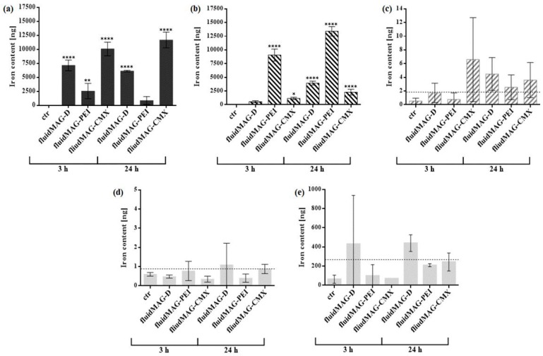
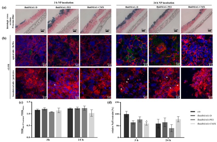
Magnetic nanoparticles are interesting tools for biomedicine. Before application, critical prerequisites have to be fulfilled. An important issue is the contact and interaction with biological barriers such as the blood-placenta barrier. In order to study these processes in detail, suitable in vitro models are needed. For that purpose a blood-placenta barrier model based on the trophoblast-like cell line BeWo and primary placenta-derived pericytes was established. This model was characterized by molecular permeability, transepithelial electrical resistance and cell-cell-contact markers. Superparamagnetic iron oxide nanoparticles (SPIONs) with cationic, anionic or neutral surface charge were applied. The localization of the nanoparticles within the cells was illustrated by histochemistry. The time-dependent passage of the nanoparticles through the BeWo/pericyte barrier was measured by magnetic particle spectroscopy and atomic absorption spectroscopy. Cationically coated SPIONs exhibited the most extensive interaction with the BeWo cells and remained primarily in the BeWo/pericyte cell layer. In contrast, SPIONs with neutral and anionic surface charge were able to pass the cell layer to a higher extent and could be detected beyond the barrier after 24 h. This study showed that the mode of SPION interaction with and passage through the in vitro blood-placenta barrier model depends on the surface charge and the duration of treatment.
磁性纳米颗粒是生物医学领域中引人关注的工具。在应用之前,必须满足关键的先决条件。一个重要问题是与生物屏障(如血胎盘屏障)的接触和相互作用。为了详细研究这些过程,需要合适的体外模型。为此,建立了一种基于滋养层样细胞系BeWo和原代胎盘来源周细胞的血胎盘屏障模型。该模型通过分子通透性、跨上皮电阻和细胞间接触标志物进行表征。应用了具有阳离子、阴离子或中性表面电荷的超顺磁性氧化铁纳米颗粒(SPIONs)。通过组织化学展示了纳米颗粒在细胞内的定位。通过磁性颗粒光谱法和原子吸收光谱法测量了纳米颗粒随时间穿过BeWo/周细胞屏障的情况。阳离子涂层的SPIONs与BeWo细胞表现出最广泛的相互作用,并且主要保留在BeWo/周细胞层中。相比之下,具有中性和阴离子表面电荷的SPIONs能够更大程度地穿过细胞层,并且在24小时后可以在屏障之外检测到。这项研究表明,SPIONs与体外血胎盘屏障模型的相互作用方式及其穿过该模型的情况取决于表面电荷和处理持续时间。