Center for the Developing Brain, Child Mind Institute, New York, NY 10022, USA; Center for Biomedical Imaging and Neuromodulation, Nathan Kline Institute for Psychiatric Research, Orangeburg, NY 10962, USA.
Center for Biomedical Imaging and Neuromodulation, Nathan Kline Institute for Psychiatric Research, Orangeburg, NY 10962, USA.
Cell Rep. 2018 Apr 10;23(2):429-441. doi: 10.1016/j.celrep.2018.03.049.
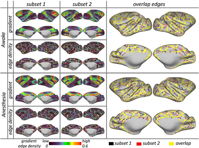
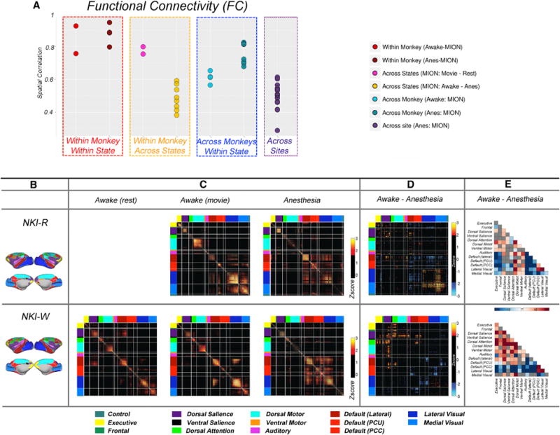
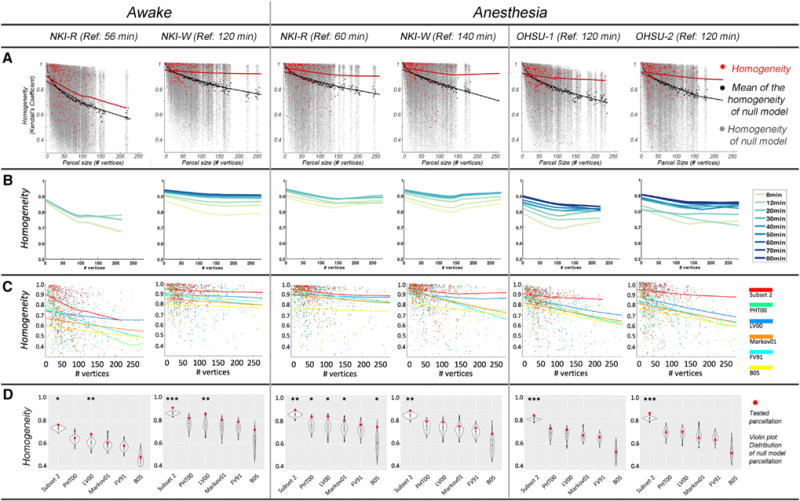
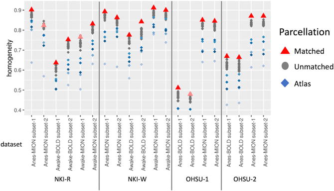
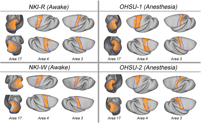
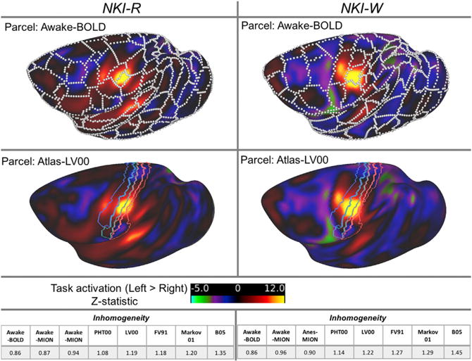
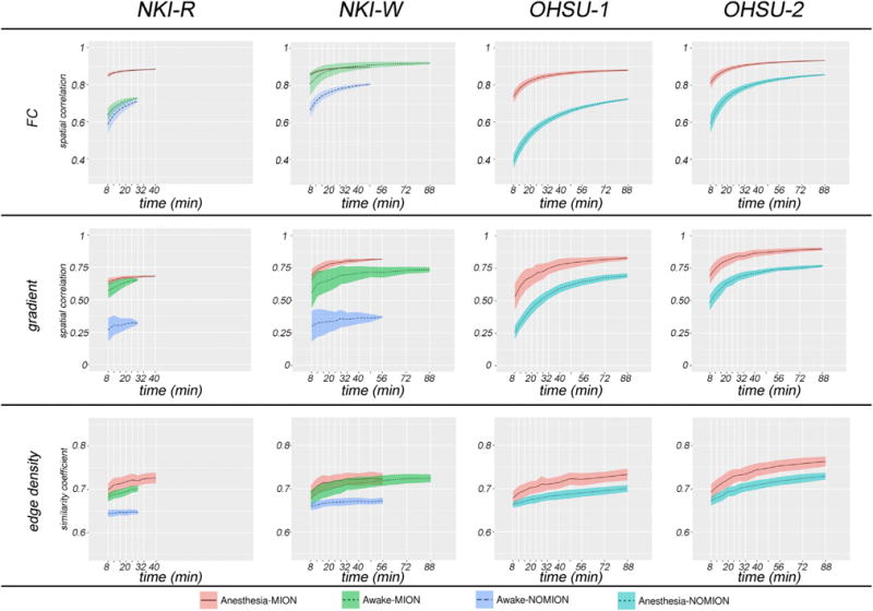
Complementing long-standing traditions centered on histology, fMRI approaches are rapidly maturing in delineating brain areal organization at the macroscale. The non-human primate (NHP) provides the opportunity to overcome critical barriers in translational research. Here, we establish the data requirements for achieving reproducible and internally valid parcellations in individuals. We demonstrate that functional boundaries serve as a functional fingerprint of the individual animals and can be achieved under anesthesia or awake conditions (rest, naturalistic viewing), though differences between awake and anesthetized states precluded the detection of individual differences across states. Comparison of awake and anesthetized states suggested a more nuanced picture of changes in connectivity for higher-order association areas, as well as visual and motor cortex. These results establish feasibility and data requirements for the generation of reproducible individual-specific parcellations in NHPs, provide insights into the impact of scan state, and motivate efforts toward harmonizing protocols.
在以组织学为中心的悠久传统基础上,功能磁共振成像(fMRI)方法在描绘宏观尺度的大脑区域组织方面迅速成熟。非人类灵长类动物(NHP)为克服转化研究中的关键障碍提供了机会。在这里,我们确定了在个体中实现可重复和内部有效的分割所需的数据要求。我们证明功能边界是个体动物的功能指纹,可以在麻醉或清醒状态下实现(休息、自然观看),尽管麻醉和清醒状态之间的差异排除了在不同状态下检测个体差异的可能性。对清醒和麻醉状态的比较表明,对于高级联合区域以及视觉和运动皮层,连接性的变化呈现出更为微妙的情况。这些结果确立了在 NHP 中生成可重复的个体特定分割的可行性和数据要求,为扫描状态的影响提供了深入了解,并促使人们努力协调方案。