• 文献检索
  • 文档翻译
  • 深度研究
  • 学术资讯
  • Suppr Zotero 插件Zotero 插件
  • 邀请有礼
  • 套餐&价格
  • 历史记录
应用&插件
Suppr Zotero 插件Zotero 插件浏览器插件Mac 客户端Windows 客户端微信小程序
定价
高级版会员购买积分包购买API积分包
服务
文献检索文档翻译深度研究API 文档MCP 服务
关于我们
关于 Suppr公司介绍联系我们用户协议隐私条款
关注我们

Suppr 超能文献

核心技术专利:CN118964589B侵权必究
粤ICP备2023148730 号-1Suppr @ 2026

文献检索

告别复杂PubMed语法,用中文像聊天一样搜索,搜遍4000万医学文献。AI智能推荐,让科研检索更轻松。

立即免费搜索

文件翻译

保留排版,准确专业,支持PDF/Word/PPT等文件格式,支持 12+语言互译。

免费翻译文档

深度研究

AI帮你快速写综述,25分钟生成高质量综述,智能提取关键信息,辅助科研写作。

立即免费体验

光学相干断层扫描血管造影术(OCTA)对中国近视人群视网膜神经纤维层和眼底血管的比较

The Comparison of Regional RNFL and Fundus Vasculature by OCTA in Chinese Myopia Population.

作者信息

Li Yuanjun, Miara Hamza, Ouyang Pingbo, Jiang Bing

机构信息

Department of Ophthalmology, Hunan Clinical Research Center of Ophthalmic Disease, The Second Xiangya Hospital, Central South University, Changsha, Hunan, China.

Department of Ophthalmology, Guangdong Provincial Key Laboratory of Malignant Tumor Epigenetics and Gene Regulation, The Sun Yat-sen Memorial Hospital, Sun Yat-sen University, Guangzhou, Guangdong, China.

出版信息

J Ophthalmol. 2018 Jan 31;2018:3490962. doi: 10.1155/2018/3490962. eCollection 2018.

DOI:10.1155/2018/3490962
PMID:29651341
原文链接:https://pmc.ncbi.nlm.nih.gov/articles/PMC5830980/
Abstract

PURPOSE

To determine the correlations between peripapillary vessel density, retinal nerve fibre layer (RNFL) thickness, and myopic indices at retina quadrants with optical coherence tomography angiography (OCTA) in Chinese.

METHODS

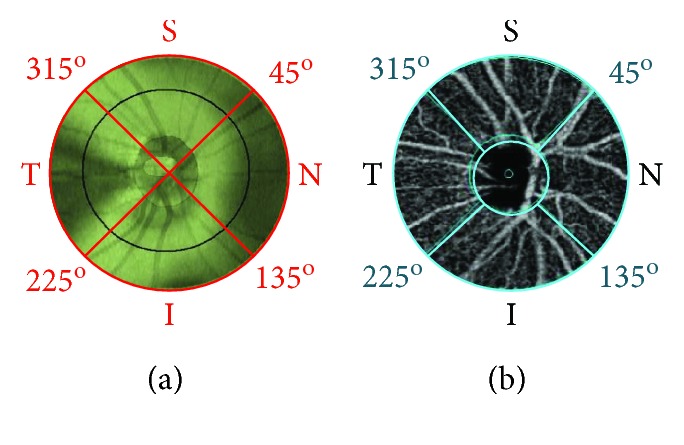
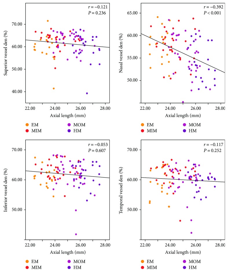
Fifty-six subjects with a mean spherical equivalent (MSE) of -3.63 ± 0.29 D were included. Peripapillary RNFL thickness and retinal vessel density in four sectors (superior, nasal, inferior, and temporal quadrants) were determined by OCTA, and correlations of the main outcomes were analyzed.

RESULTS

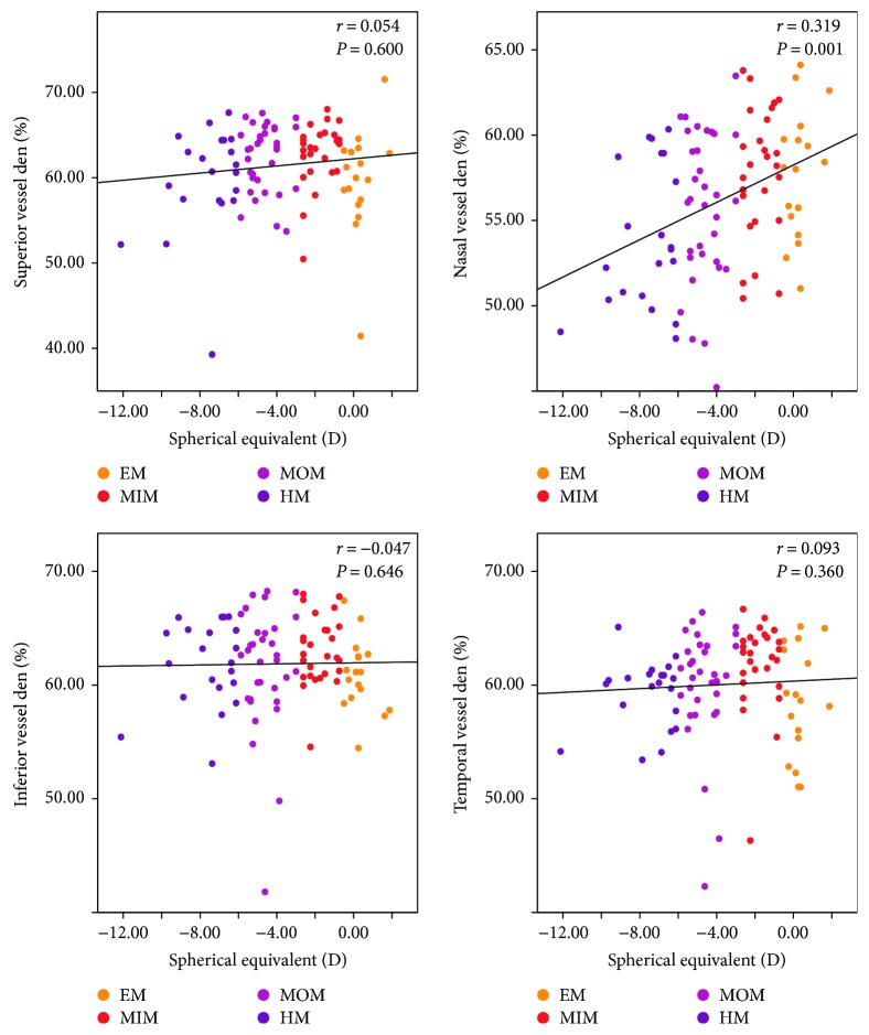
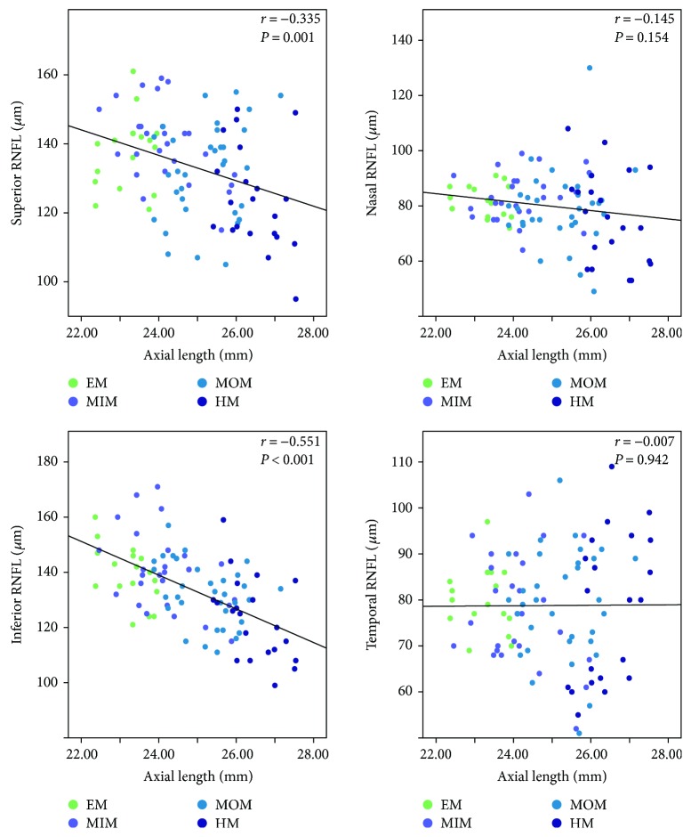
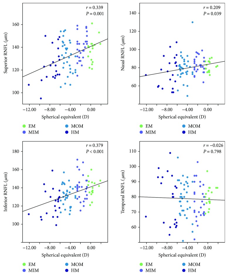
Negative correlations were found between the peripapillary RNFL thickness and axial length (AL) at superior ( = -0.335, = 0.001) and inferior ( = -0.551, < 0.001) quadrants. There was a significant positive correlation with spherical equivalent (SE) at the corresponding quadrants as well as at the nasal quadrant ( = 0.339, = 0.001; = 0.379, < 0.001; and = 0.209, = 0.039, resp.). Peripapillary retinal vessel density was also negatively correlated with AL at the nasal quadrant ( = -0.392, < 0.001), and only at the nasal quadrant, there was a positive correlation between the peripapillary vessel density and SE ( = 0.319, = 0.001).

CONCLUSIONS

The degree of myopia and elongation of AL were negatively correlated with peripapillary RNFL thickness at superior and inferior quadrants and with peripapillary retinal vessel density at the nasal quadrant.

摘要

目的

运用光学相干断层扫描血管造影(OCTA)技术,在中国人群中确定视乳头周围血管密度、视网膜神经纤维层(RNFL)厚度与视网膜象限近视指标之间的相关性。

方法

纳入56名平均球镜等效度数(MSE)为-3.63±0.29D的受试者。通过OCTA测定四个象限(上方、鼻侧、下方和颞侧)的视乳头周围RNFL厚度和视网膜血管密度,并分析主要结果的相关性。

结果

上方象限(r=-0.335,P=0.001)和下方象限(r=-0.551,P<0.001)的视乳头周围RNFL厚度与眼轴长度(AL)呈负相关。在相应象限以及鼻侧象限,视乳头周围RNFL厚度与球镜等效度数(SE)呈显著正相关(分别为r=0.339,P=0.001;r=0.379,P<0.001;r=0.209,P=0.039)。鼻侧象限的视乳头周围视网膜血管密度也与AL呈负相关(r=-0.392,P<0.001),且仅在鼻侧象限,视乳头周围血管密度与SE呈正相关(r=0.319,P=0.001)。

结论

近视程度和AL延长与上方和下方象限的视乳头周围RNFL厚度以及鼻侧象限的视乳头周围视网膜血管密度呈负相关。

https://cdn.ncbi.nlm.nih.gov/pmc/blobs/aea2/5830980/a4836c7df9d5/JOPH2018-3490962.005.jpg
https://cdn.ncbi.nlm.nih.gov/pmc/blobs/aea2/5830980/8249d1ea1c81/JOPH2018-3490962.001.jpg
https://cdn.ncbi.nlm.nih.gov/pmc/blobs/aea2/5830980/cc25f7d410dc/JOPH2018-3490962.002.jpg
https://cdn.ncbi.nlm.nih.gov/pmc/blobs/aea2/5830980/bbb21f161438/JOPH2018-3490962.003.jpg
https://cdn.ncbi.nlm.nih.gov/pmc/blobs/aea2/5830980/1e09a0a1a005/JOPH2018-3490962.004.jpg
https://cdn.ncbi.nlm.nih.gov/pmc/blobs/aea2/5830980/a4836c7df9d5/JOPH2018-3490962.005.jpg

相似文献

1
The Comparison of Regional RNFL and Fundus Vasculature by OCTA in Chinese Myopia Population.光学相干断层扫描血管造影术(OCTA)对中国近视人群视网膜神经纤维层和眼底血管的比较
J Ophthalmol. 2018 Jan 31;2018:3490962. doi: 10.1155/2018/3490962. eCollection 2018.
2
Topographic Quadrant Analysis of Peripapillary Superficial Microvasculature in Optic Disc Drusen.视盘小凹周围浅表微血管的象限分析
Front Neurol. 2021 May 19;12:666359. doi: 10.3389/fneur.2021.666359. eCollection 2021.
3
Evaluation of peripapillary retinal nerve fiber layer thickness of myopic and hyperopic patients: a controlled study by Stratus optical coherence tomography.近视与远视患者的视盘周围视网膜神经纤维层厚度评估:Stratus 光学相干断层扫描的对照研究。
Curr Eye Res. 2013 Jan;38(1):102-7. doi: 10.3109/02713683.2012.715714. Epub 2012 Aug 22.
4
Macular and peripapillary vessel density in myopic eyes of young Chinese adults.中国年轻近视人群的黄斑区及视盘周围血管密度。
Clin Exp Optom. 2020 Nov;103(6):830-837. doi: 10.1111/cxo.13047. Epub 2020 Feb 12.
5
Peripapillary retinal nerve fiber layer thickness distribution in Chinese with myopia measured by 3D-optical coherence tomography.采用三维光学相干断层扫描测量的中国近视人群视乳头周围视网膜神经纤维层厚度分布
Int J Ophthalmol. 2013 Oct 18;6(5):626-31. doi: 10.3980/j.issn.2222-3959.2013.05.13. eCollection 2013.
6
Peripapillary vessel density measurement of quadrant and clock-hour sectors in primary angle closure glaucoma using optical coherence tomography angiography.应用光学相干断层扫描血管造影术测量原发性闭角型青光眼象限和时钟小时区的视盘周围血管密度。
BMC Ophthalmol. 2021 Sep 9;21(1):328. doi: 10.1186/s12886-021-02093-0.
7
Peripapillary Retinal Nerve Fiber Layer (RNFL) Thickness Measurements by Topcon SD-OCT in Myopic Patients.托普康频域光学相干断层扫描技术测量近视患者视乳头周围视网膜神经纤维层厚度
J Coll Physicians Surg Pak. 2018 Jan;28(1):26-30. doi: 10.29271/jcpsp.2018.01.26.
8
Nomogram of peripapillary retinal nerve fiber layer thickness in myopic eyes of north Indian population.北印度人群近视眼中的视盘周围视网膜神经纤维层厚度的诺莫图。
Indian J Ophthalmol. 2022 Feb;70(2):458-464. doi: 10.4103/ijo.IJO_767_21.
9
Reliability of Vessel Density Measurements in the Peripapillary Retina and Correlation with Retinal Nerve Fiber Layer Thickness in Healthy Subjects Using Optical Coherence Tomography Angiography.使用光学相干断层扫描血管造影术测量健康受试者视乳头周围视网膜血管密度的可靠性及其与视网膜神经纤维层厚度的相关性
Ophthalmologica. 2018;240(4):183-190. doi: 10.1159/000485957. Epub 2018 Apr 25.
10
The association between macular thickness and peripapillary retinal nerve fiber layer thickness in Chinese children.中国儿童黄斑厚度与视乳头周围视网膜神经纤维层厚度之间的关联。
Medicine (Baltimore). 2015 Feb;94(8):e567. doi: 10.1097/MD.0000000000000567.

引用本文的文献

1
Effects of Chinese Acupoint Eye Exercise on Ocular Biological Parameters.中医眼保健操对眼部生物学参数的影响。
Transl Vis Sci Technol. 2025 Jul 1;14(7):20. doi: 10.1167/tvst.14.7.20.
2
Interocular comparison of peripapillary retinal nerve fiber layer thickness and vasculature in non-pathological myopia with anisometropia.非病理性屈光参差性近视患者视乳头周围视网膜神经纤维层厚度和血管系统的双眼比较
Graefes Arch Clin Exp Ophthalmol. 2025 Apr 8. doi: 10.1007/s00417-025-06826-5.
3
Evidence of vascular involvement in myopia: a review.近视中血管受累的证据:综述

本文引用的文献

1
Evaluation of Optic Nerve Head Parameters and Retinal Nerve Fiber Layer Thickness in Axial Myopia Using SD OCT.使用频域光学相干断层扫描技术评估轴性近视患者的视神经乳头参数和视网膜神经纤维层厚度
Ophthalmol Ther. 2017 Dec;6(2):335-341. doi: 10.1007/s40123-017-0095-5. Epub 2017 Jun 5.
2
Exploration of peripapillary vessel density in highly myopic eyes with peripapillary intrachoroidal cavitation and its relationship with ocular parameters using optical coherence tomography angiography.应用光相干断层扫描血管造影术对伴有脉络膜视网膜神经上皮层下腔的高度近视眼视盘周围血管密度的研究及其与眼参数的关系。
Clin Exp Ophthalmol. 2017 Dec;45(9):884-893. doi: 10.1111/ceo.12986. Epub 2017 Jun 15.
3
Front Med (Lausanne). 2023 May 18;10:1112996. doi: 10.3389/fmed.2023.1112996. eCollection 2023.
4
Prospective clinical study of retinal microvascular alteration after ICL implantation.ICL植入术后视网膜微血管改变的前瞻性临床研究
Front Cell Dev Biol. 2023 Jan 19;11:1115822. doi: 10.3389/fcell.2023.1115822. eCollection 2023.
5
Analysis of Microcirculation Changes in the Macular Area and Para-Optic Disk Region After Implantable Collamer Lens Implantation in Patients With High Myopia.高度近视患者植入可植入式胶原晶状体后黄斑区和视盘旁区域微循环变化的分析
Front Neurosci. 2022 May 19;16:867463. doi: 10.3389/fnins.2022.867463. eCollection 2022.
6
An Evaluation of Choroidal and Retinal Nerve Fiber Layer Thicknesses Using SD-OCT in Children with Childhood IgA Vasculitis.使用频域光学相干断层扫描评估儿童IgA血管炎患儿的脉络膜和视网膜神经纤维层厚度
Diagnostics (Basel). 2022 Apr 5;12(4):901. doi: 10.3390/diagnostics12040901.
7
Vessel Density in the Macular and Peripapillary Areas in Preperimetric Glaucoma to Various Stages of Primary Open-Angle Glaucoma in Taiwan.台湾青光眼前期黄斑区和视乳头周围区域血管密度与原发性开角型青光眼各阶段的关系
J Clin Med. 2021 Nov 23;10(23):5490. doi: 10.3390/jcm10235490.
8
Macular Vessel Density Changes in Young Adults With High Myopia: A Longitudinal Study.高度近视青年成人黄斑区血管密度变化:一项纵向研究
Front Med (Lausanne). 2021 Jun 8;8:648644. doi: 10.3389/fmed.2021.648644. eCollection 2021.
9
Quantitative assessment and determinants of the papillary microvasculature in healthy subjects.健康受试者乳头微血管的定量评估及其决定因素。
BMC Ophthalmol. 2021 Mar 20;21(1):140. doi: 10.1186/s12886-021-01896-5.
10
Macular vessel density and foveal avascular zone parameters in patients after acute primary angle closure determined by OCT angiography.OCT 血管造影术评估急性原发性闭角型青光眼患者的黄斑血管密度和中心无血管区参数。
Sci Rep. 2020 Oct 30;10(1):18717. doi: 10.1038/s41598-020-73223-9.
Macular Ganglion Cell and Retinal Nerve Fiber Layer Thickness in Children With Refractive Errors-An Optical Coherence Tomography Study.
屈光不正儿童的黄斑神经节细胞和视网膜神经纤维层厚度——一项光学相干断层扫描研究
J Glaucoma. 2017 Jul;26(7):619-625. doi: 10.1097/IJG.0000000000000683.
4
Regional vascular density-visual field sensitivity relationship in glaucoma according to disease severity.青光眼患者中根据疾病严重程度的局部血管密度与视野敏感度关系
Br J Ophthalmol. 2017 Dec;101(12):1666-1672. doi: 10.1136/bjophthalmol-2017-310180. Epub 2017 Apr 21.
5
Quantitative OCT Angiography of the Retinal Microvasculature and the Choriocapillaris in Myopic Eyes.近视眼中视网膜微血管和脉络膜毛细血管的定量光学相干断层扫描血管造影
Invest Ophthalmol Vis Sci. 2017 Apr 1;58(4):2063-2069. doi: 10.1167/iovs.16-21289.
6
Ganglion cell-inner plexiform layer and retinal nerve fiber layer thickness according to myopia and optic disc area: a quantitative and three-dimensional analysis.根据近视和视盘面积分析神经节细胞-内网状层及视网膜神经纤维层厚度:定量与三维分析
BMC Ophthalmol. 2017 Mar 11;17(1):22. doi: 10.1186/s12886-017-0419-1.
7
Reduced Macular Vascular Density in Myopic Eyes.近视眼中黄斑区血管密度降低。
Chin Med J (Engl). 2017 Feb 20;130(4):445-451. doi: 10.4103/0366-6999.199844.
8
Retinal Microvasculature Alteration in High Myopia.高度近视患者视网膜微血管改变
Invest Ophthalmol Vis Sci. 2016 Nov 1;57(14):6020-6030. doi: 10.1167/iovs.16-19542.
9
Retinal Microvascular Network and Microcirculation Assessments in High Myopia.高度近视患者视网膜微血管网络及微循环评估
Am J Ophthalmol. 2017 Feb;174:56-67. doi: 10.1016/j.ajo.2016.10.018. Epub 2016 Nov 4.
10
Optical coherence tomography angiography: an overview of the technology and an assessment of applications for clinical research.光学相干断层扫描血管造影术:技术概述及临床研究应用评估
Br J Ophthalmol. 2017 Jan;101(1):16-20. doi: 10.1136/bjophthalmol-2016-309389. Epub 2016 Oct 4.