Aboushady Iman M, Salem Zeinab A, Sabry Dina, Mohamed Abbas
MD, MS, Lecturer of oral biology, Department of Oral Biology, Faculty of Oral and Dental Medicine, Cairo University.
MD, MS, Professor of Medical Biochemistry and Molecular Biology, Department of Medical biochemistry and molecular biology, Faculty of medicine, Cairo University.
J Clin Exp Dent. 2018 Jan 1;10(1):e7-e13. doi: 10.4317/jced.53957. eCollection 2018 Jan.
Mesenchymal stem cells (MSCs) can regenerate missing tissues and treat diseases. Hence, the current work aimed to compare the proliferation rate and the osteogenic differentiation potential of bone marrow MSCs (BMSCs), gingival MSCs (GMSCs) and submandibular MSCs (SMSCs).
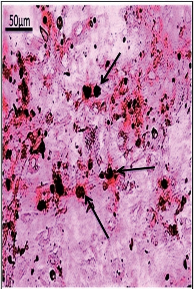
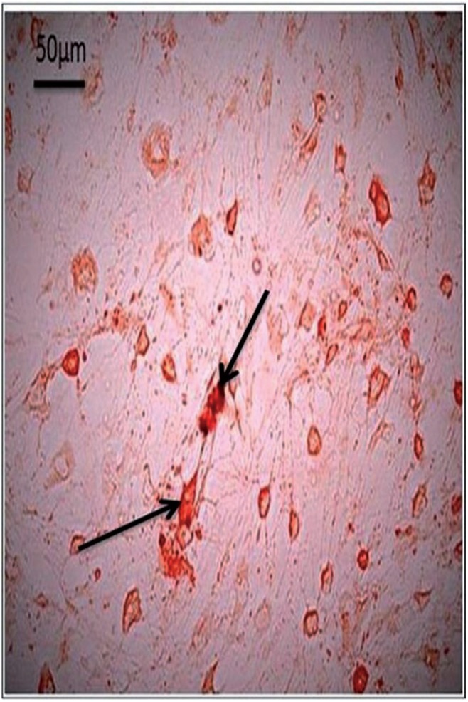
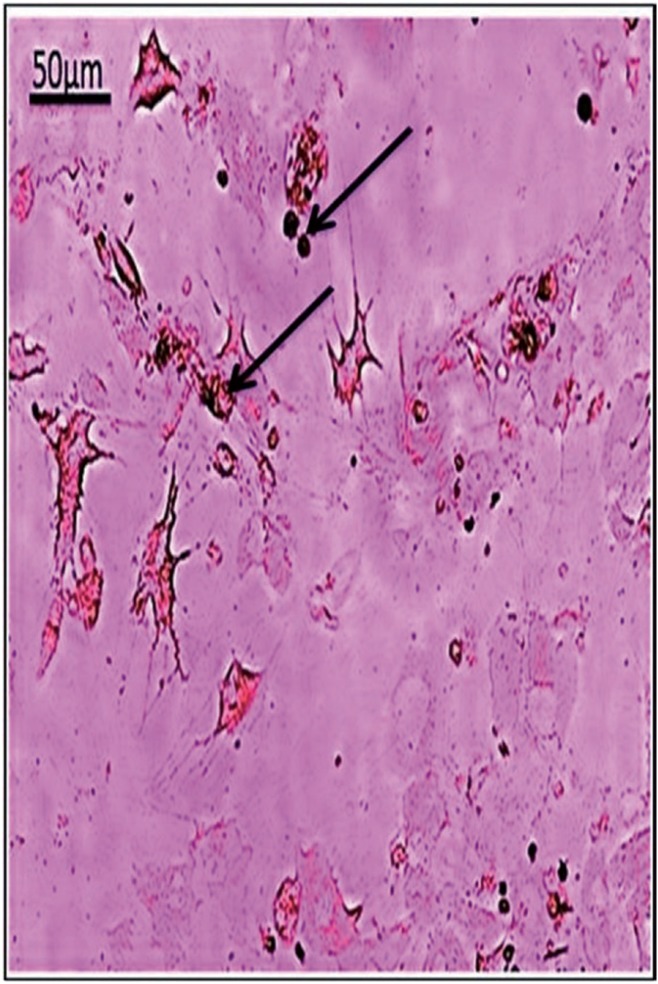
MSCs derived from bone marrow, gingiva and submandibular salivary gland were isolated and cultured from rats. The proliferation capacity was judged by MTT proliferation Assay. Osteogenic differentiation was assessed by Alzarin red stain and quantitative RT-PCR was performed for Runx-2 and MMP-13.
The highest significant proliferation was estimated in the BMSCs compared to GMSCs and SMSCs (-value was < 0.01). All studied cell types formed mineralized nodules as stained with Alizarin Red stain at the 3rd passage of differentiation. However, BMSCs seemed to generate the highest level of mineralization compared to GMSCs and SMSCs. RT-PCR revealed that the expression of Runx-2 and MMP-13 mRNAs was significantly increased in the BMSCs compared to GMSCs and SMSCs (-value was < 0.01).
BMSCs displayed maximum osteogenesis results followed by the GMSCs and lastly by the SGSCs. Thus, it could be recommended that GMSCs can be used as a second choice after BMSCs when bone tissue reconstruction is needed. Mesenchymal stem cells, osteogenic differentiation, Runx-2, MMP-13.
间充质干细胞(MSCs)可再生缺失组织并治疗疾病。因此,当前研究旨在比较骨髓间充质干细胞(BMSCs)、牙龈间充质干细胞(GMSCs)和下颌下间充质干细胞(SMSCs)的增殖率和成骨分化潜能。
从大鼠中分离并培养源自骨髓、牙龈和下颌下唾液腺的间充质干细胞。通过MTT增殖试验判断增殖能力。通过茜素红染色评估成骨分化,并对Runx-2和MMP-13进行定量逆转录聚合酶链反应(RT-PCR)。
与GMSCs和SMSCs相比,BMSCs的增殖最为显著(P值<0.01)。在分化的第3代,所有研究的细胞类型经茜素红染色均形成矿化结节。然而,与GMSCs和SMSCs相比,BMSCs似乎产生的矿化水平最高。RT-PCR显示,与GMSCs和SMSCs相比,BMSCs中Runx-2和MMP-13 mRNA的表达显著增加(P值<0.01)。
BMSCs的成骨效果最佳,其次是GMSCs,最后是SMSCs。因此,当需要进行骨组织重建时,建议在BMSCs之后可将GMSCs作为第二选择。间充质干细胞、成骨分化、Runx-2、MMP-13。