Chen Wei, Liu Boqiang, Peng Suting, Sun Jiawei, Qiao Xu
Department of Biomedical Engineering, School of Control Science and Engineering, Shandong University, Shandong, China.
Int J Biomed Imaging. 2018 May 8;2018:2512037. doi: 10.1155/2018/2512037. eCollection 2018.
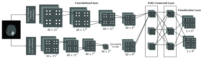
Gliomas are the most common primary brain tumors, and the objective grading is of great importance for treatment. This paper presents an automatic computer-aided diagnosis of gliomas that combines automatic segmentation and radiomics, which can improve the diagnostic ability. The MRI data containing 220 high-grade gliomas and 54 low-grade gliomas are used to evaluate our system. A multiscale 3D convolutional neural network is trained to segment whole tumor regions. A wide range of radiomic features including first-order features, shape features, and texture features is extracted. By using support vector machines with recursive feature elimination for feature selection, a CAD system that has an extreme gradient boosting classifier with a 5-fold cross-validation is constructed for the grading of gliomas. Our CAD system is highly effective for the grading of gliomas with an accuracy of 91.27%, a weighted macroprecision of 91.27%, a weighted macrorecall of 91.27%, and a weighted macro-1 score of 90.64%. This demonstrates that the proposed CAD system can assist radiologists for high accurate grading of gliomas and has the potential for clinical applications.
胶质瘤是最常见的原发性脑肿瘤,客观分级对治疗至关重要。本文提出了一种结合自动分割和放射组学的胶质瘤自动计算机辅助诊断方法,可提高诊断能力。使用包含220例高级别胶质瘤和54例低级别胶质瘤的MRI数据来评估我们的系统。训练一个多尺度3D卷积神经网络来分割整个肿瘤区域。提取包括一阶特征、形状特征和纹理特征在内的广泛的放射组学特征。通过使用带有递归特征消除的支持向量机进行特征选择,构建了一个具有极端梯度提升分类器并采用五折交叉验证的CAD系统用于胶质瘤分级。我们的CAD系统对胶质瘤分级非常有效,准确率为91.27%,加权宏观精度为91.27%,加权宏观召回率为91.27%,加权宏观F1分数为90.64%。这表明所提出的CAD系统可以协助放射科医生对胶质瘤进行高精度分级,并具有临床应用潜力。