• 文献检索
  • 文档翻译
  • 深度研究
  • 学术资讯
  • Suppr Zotero 插件Zotero 插件
  • 邀请有礼
  • 套餐&价格
  • 历史记录
应用&插件
Suppr Zotero 插件Zotero 插件浏览器插件Mac 客户端Windows 客户端微信小程序
定价
高级版会员购买积分包购买API积分包
服务
文献检索文档翻译深度研究API 文档MCP 服务
关于我们
关于 Suppr公司介绍联系我们用户协议隐私条款
关注我们

Suppr 超能文献

核心技术专利:CN118964589B侵权必究
粤ICP备2023148730 号-1Suppr @ 2026

文献检索

告别复杂PubMed语法,用中文像聊天一样搜索,搜遍4000万医学文献。AI智能推荐,让科研检索更轻松。

立即免费搜索

文件翻译

保留排版,准确专业,支持PDF/Word/PPT等文件格式,支持 12+语言互译。

免费翻译文档

深度研究

AI帮你快速写综述,25分钟生成高质量综述,智能提取关键信息,辅助科研写作。

立即免费体验

使用深度卷积神经网络进行精确的脑肿瘤检测。

Accurate brain tumor detection using deep convolutional neural network.

作者信息

Khan Md Saikat Islam, Rahman Anichur, Debnath Tanoy, Karim Md Razaul, Nasir Mostofa Kamal, Band Shahab S, Mosavi Amir, Dehzangi Iman

机构信息

Department of CSE, Mawlana Bhashani Science and Technology University, Tangail, Bangladesh.

Department of CSE, National Institute of Textile Engineering and Research (NITER), Constituent Institute of the University of Dhaka, Savar, Dhaka 1350, Bangladesh.

出版信息

Comput Struct Biotechnol J. 2022 Aug 27;20:4733-4745. doi: 10.1016/j.csbj.2022.08.039. eCollection 2022.

DOI:10.1016/j.csbj.2022.08.039
PMID:36147663
原文链接:https://pmc.ncbi.nlm.nih.gov/articles/PMC9468505/
Abstract

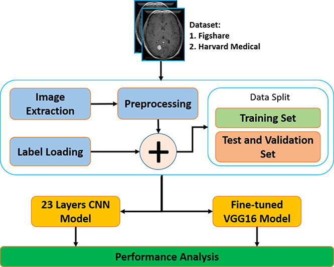
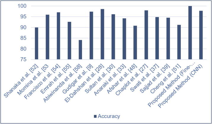
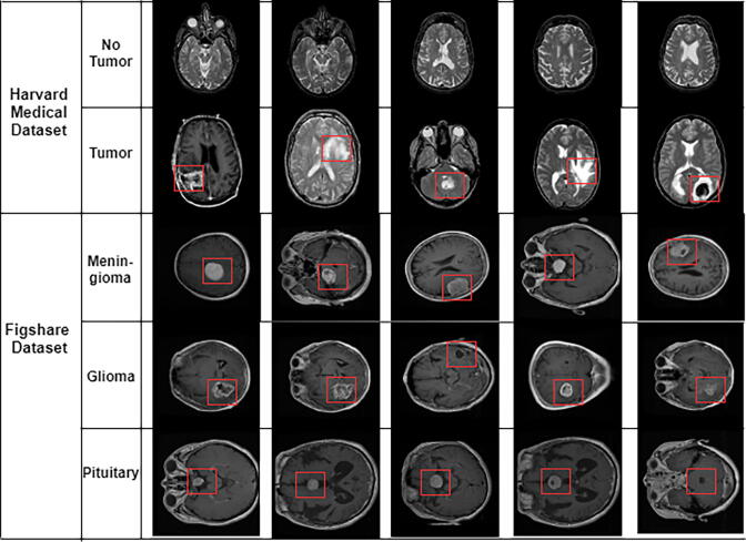
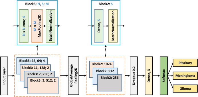
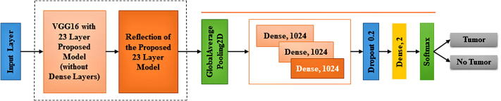
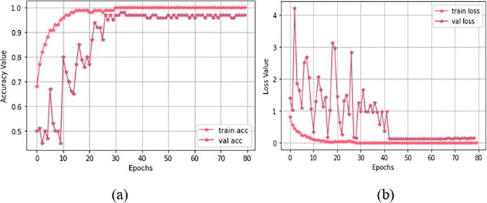
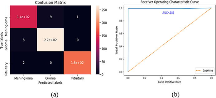
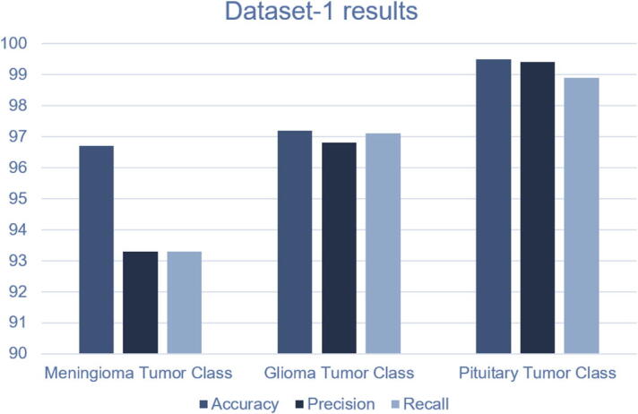
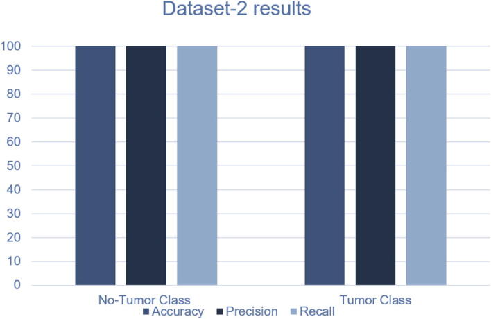
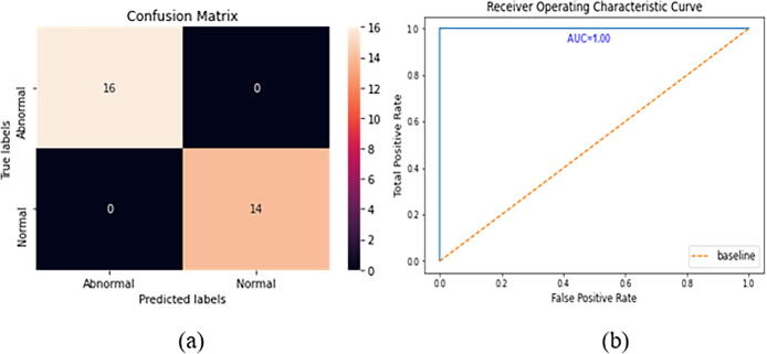
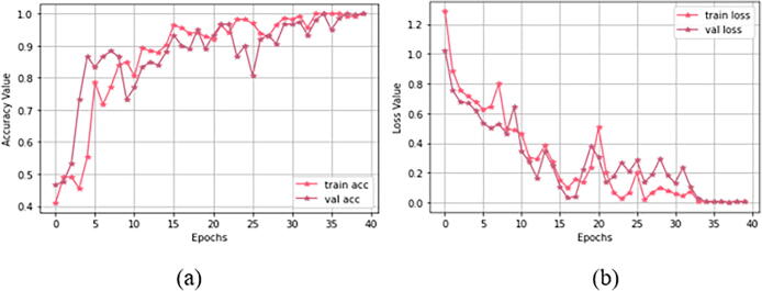
Detection and Classification of a brain tumor is an important step to better understanding its mechanism. Magnetic Reasoning Imaging (MRI) is an experimental medical imaging technique that helps the radiologist find the tumor region. However, it is a time taking process and requires expertise to test the MRI images, manually. Nowadays, the advancement of Computer-assisted Diagnosis (CAD), machine learning, and deep learning in specific allow the radiologist to more reliably identify brain tumors. The traditional machine learning methods used to tackle this problem require a handcrafted feature for classification purposes. Whereas deep learning methods can be designed in a way to not require any handcrafted feature extraction while achieving accurate classification results. This paper proposes two deep learning models to identify both binary (normal and abnormal) and multiclass (meningioma, glioma, and pituitary) brain tumors. We use two publicly available datasets that include 3064 and 152 MRI images, respectively. To build our models, we first apply a 23-layers convolution neural network (CNN) to the first dataset since there is a large number of MRI images for the training purpose. However, when dealing with limited volumes of data, which is the case in the second dataset, our proposed "23-layers CNN" architecture faces overfitting problem. To address this issue, we use transfer learning and combine VGG16 architecture along with the reflection of our proposed "23 layers CNN" architecture. Finally, we compare our proposed models with those reported in the literature. Our experimental results indicate that our models achieve up to 97.8% and 100% classification accuracy for our employed datasets, respectively, exceeding all other state-of-the-art models. Our proposed models, employed datasets, and all the source codes are publicly available at: (https://github.com/saikat15010/Brain-Tumor-Detection).

摘要

脑肿瘤的检测与分类是更好地理解其发病机制的重要一步。磁共振成像(MRI)是一种实验性医学成像技术,可帮助放射科医生找到肿瘤区域。然而,这是一个耗时的过程,并且需要专业知识来手动检测MRI图像。如今,计算机辅助诊断(CAD)、机器学习和深度学习的发展尤其使放射科医生能够更可靠地识别脑肿瘤。用于解决此问题的传统机器学习方法需要手工制作的特征用于分类目的。而深度学习方法可以设计成无需任何手工特征提取就能获得准确的分类结果。本文提出了两种深度学习模型来识别二元(正常和异常)和多类(脑膜瘤、胶质瘤和垂体瘤)脑肿瘤。我们使用了两个公开可用的数据集,分别包含3064张和152张MRI图像。为了构建我们的模型,由于有大量用于训练目的的MRI图像,我们首先将一个23层卷积神经网络(CNN)应用于第一个数据集。然而,在处理数据量有限的情况时,也就是第二个数据集的情况,我们提出的“23层CNN”架构面临过拟合问题。为了解决这个问题,我们使用迁移学习并将VGG16架构与我们提出的“23层CNN”架构的改进相结合。最后,我们将我们提出的模型与文献中报道的模型进行比较。我们的实验结果表明,我们的模型在所使用的数据集上分别达到了高达97.8%和100%的分类准确率,超过了所有其他现有最先进的模型。我们提出的模型、所使用的数据集以及所有源代码均可在以下网址公开获取:(https://github.com/saikat15010/Brain-Tumor-Detection)

https://cdn.ncbi.nlm.nih.gov/pmc/blobs/1f25/9468505/1f30aa78141b/gr15.jpg
https://cdn.ncbi.nlm.nih.gov/pmc/blobs/1f25/9468505/dfefcd0f391f/ga1.jpg
https://cdn.ncbi.nlm.nih.gov/pmc/blobs/1f25/9468505/d60fe69f3bca/gr1.jpg
https://cdn.ncbi.nlm.nih.gov/pmc/blobs/1f25/9468505/a8619fc5aaab/gr2.jpg
https://cdn.ncbi.nlm.nih.gov/pmc/blobs/1f25/9468505/8a4ecad15ac4/gr3.jpg
https://cdn.ncbi.nlm.nih.gov/pmc/blobs/1f25/9468505/12ad7e13b80c/gr4.jpg
https://cdn.ncbi.nlm.nih.gov/pmc/blobs/1f25/9468505/003b214dc037/gr5.jpg
https://cdn.ncbi.nlm.nih.gov/pmc/blobs/1f25/9468505/952842672254/gr6.jpg
https://cdn.ncbi.nlm.nih.gov/pmc/blobs/1f25/9468505/c0deaca01020/gr7.jpg
https://cdn.ncbi.nlm.nih.gov/pmc/blobs/1f25/9468505/9ea82f791c3d/gr8.jpg
https://cdn.ncbi.nlm.nih.gov/pmc/blobs/1f25/9468505/ee76e50afe0d/gr9.jpg
https://cdn.ncbi.nlm.nih.gov/pmc/blobs/1f25/9468505/e334bcd1fce7/gr10.jpg
https://cdn.ncbi.nlm.nih.gov/pmc/blobs/1f25/9468505/825c8aca663e/gr11.jpg
https://cdn.ncbi.nlm.nih.gov/pmc/blobs/1f25/9468505/f1d6e396da5a/gr12.jpg
https://cdn.ncbi.nlm.nih.gov/pmc/blobs/1f25/9468505/af3572257f10/gr13.jpg
https://cdn.ncbi.nlm.nih.gov/pmc/blobs/1f25/9468505/9af2818e81ca/gr14.jpg
https://cdn.ncbi.nlm.nih.gov/pmc/blobs/1f25/9468505/1f30aa78141b/gr15.jpg

相似文献

1
Accurate brain tumor detection using deep convolutional neural network.使用深度卷积神经网络进行精确的脑肿瘤检测。
Comput Struct Biotechnol J. 2022 Aug 27;20:4733-4745. doi: 10.1016/j.csbj.2022.08.039. eCollection 2022.
2
MRI-based brain tumor detection using convolutional deep learning methods and chosen machine learning techniques.基于 MRI 的脑肿瘤检测:使用卷积深度学习方法和选定的机器学习技术。
BMC Med Inform Decis Mak. 2023 Jan 23;23(1):16. doi: 10.1186/s12911-023-02114-6.
3
A Hybrid Approach Based on Deep CNN and Machine Learning Classifiers for the Tumor Segmentation and Classification in Brain MRI.基于深度卷积神经网络和机器学习分类器的脑 MRI 肿瘤分割与分类的混合方法。
Comput Math Methods Med. 2022 Aug 5;2022:6446680. doi: 10.1155/2022/6446680. eCollection 2022.
4
Tumor Diagnosis against Other Brain Diseases Using T2 MRI Brain Images and CNN Binary Classifier and DWT.使用T2加权磁共振成像脑图像、卷积神经网络二元分类器和离散小波变换进行肿瘤与其他脑部疾病的诊断
Brain Sci. 2023 Feb 17;13(2):348. doi: 10.3390/brainsci13020348.
5
Brain tumor classification for MR images using transfer learning and fine-tuning.基于迁移学习和微调的磁共振图像脑肿瘤分类。
Comput Med Imaging Graph. 2019 Jul;75:34-46. doi: 10.1016/j.compmedimag.2019.05.001. Epub 2019 May 18.
6
Integrated approach of federated learning with transfer learning for classification and diagnosis of brain tumor.联邦学习与迁移学习相结合的综合方法用于脑肿瘤的分类和诊断。
BMC Med Imaging. 2024 May 15;24(1):110. doi: 10.1186/s12880-024-01261-0.
7
An Efficient Multi-Scale Convolutional Neural Network Based Multi-Class Brain MRI Classification for SaMD.基于高效多尺度卷积神经网络的 SaMD 多类脑 MRI 分类
Tomography. 2022 Jul 26;8(4):1905-1927. doi: 10.3390/tomography8040161.
8
Brain tumour classification of magnetic resonance images using a novel CNN-based medical image analysis and detection network in comparison to VGG16.基于新型 CNN 的医学图像分析与检测网络与 VGG16 对磁共振图像进行脑部肿瘤分类比较
J Popul Ther Clin Pharmacol. 2022 Jan 26;28(2):e113-e125. doi: 10.47750/jptcp.2022.873. eCollection 2022.
9
An efficient brain tumor image classifier by combining multi-pathway cascaded deep neural network and handcrafted features in MR images.基于多通路级联深度神经网络和磁共振图像手工特征融合的脑肿瘤图像高效分类器
Med Biol Eng Comput. 2021 Aug;59(7-8):1495-1527. doi: 10.1007/s11517-021-02370-6. Epub 2021 Jun 29.
10
Deep Multi-Scale 3D Convolutional Neural Network (CNN) for MRI Gliomas Brain Tumor Classification.基于深度多尺度 3D 卷积神经网络(CNN)的 MRI 脑肿瘤胶质瘤分类。
J Digit Imaging. 2020 Aug;33(4):903-915. doi: 10.1007/s10278-020-00347-9.

引用本文的文献

1
Spinal-QDCNN: advanced feature extraction for brain tumor detection using MRI images.脊髓-QDCNN:使用MRI图像进行脑肿瘤检测的高级特征提取
Eur Spine J. 2025 Aug 9. doi: 10.1007/s00586-025-09147-7.
2
Performance Evaluation of Artificial Intelligence Techniques in the Diagnosis of Brain Tumors: A Systematic Review and Meta-Analysis.人工智能技术在脑肿瘤诊断中的性能评估:系统评价与荟萃分析
Cureus. 2025 Jul 28;17(7):e88915. doi: 10.7759/cureus.88915. eCollection 2025 Jul.
3
Hybrid Attention-Enhanced Xception and Dynamic Chaotic Whale Optimization for Brain Tumor Diagnosis.

本文引用的文献

1
Brain Tumor Detection and Classification by MRI Using Biologically Inspired Orthogonal Wavelet Transform and Deep Learning Techniques.基于生物启发正交小波变换和深度学习技术的 MRI 脑肿瘤检测与分类。
J Healthc Eng. 2022 Jan 10;2022:2693621. doi: 10.1155/2022/2693621. eCollection 2022.
2
Brain Tumor/Mass Classification Framework Using Magnetic-Resonance-Imaging-Based Isolated and Developed Transfer Deep-Learning Model.基于磁共振成像的孤立开发转移深度学习模型的脑肿瘤/肿块分类框架。
Sensors (Basel). 2022 Jan 4;22(1):372. doi: 10.3390/s22010372.
3
A Novel Deep Learning Method for Recognition and Classification of Brain Tumors from MRI Images.
用于脑肿瘤诊断的混合注意力增强型Xception和动态混沌鲸鱼优化算法
Bioengineering (Basel). 2025 Jul 9;12(7):747. doi: 10.3390/bioengineering12070747.
4
Intelligent brain tumor detection using hybrid finetuned deep transfer features and ensemble machine learning algorithms.使用混合微调深度迁移特征和集成机器学习算法的智能脑肿瘤检测
Sci Rep. 2025 Jul 4;15(1):23899. doi: 10.1038/s41598-025-08689-6.
5
A federated learning-based privacy-preserving image processing framework for brain tumor detection from CT scans.一种基于联邦学习的用于从CT扫描中检测脑肿瘤的隐私保护图像处理框架。
Sci Rep. 2025 Jul 2;15(1):23578. doi: 10.1038/s41598-025-07807-8.
6
Automated Brain Tumor Classification and Grading Using Multi-scale Graph Neural Network with Spatio-Temporal Transformer Attention Through MRI Scans.通过MRI扫描,使用具有时空Transformer注意力的多尺度图神经网络进行脑肿瘤自动分类和分级
Interdiscip Sci. 2025 Jun 5. doi: 10.1007/s12539-025-00718-2.
7
Prediction of pathological grade of oral squamous cell carcinoma and construction of prognostic model based on deep learning algorithm.基于深度学习算法的口腔鳞状细胞癌病理分级预测及预后模型构建
Discov Oncol. 2025 Jun 1;16(1):976. doi: 10.1007/s12672-025-02144-8.
8
An explainable AI-driven deep neural network for accurate breast cancer detection from histopathological and ultrasound images.一种可解释的人工智能驱动的深度神经网络,用于从组织病理学和超声图像中准确检测乳腺癌。
Sci Rep. 2025 May 20;15(1):17531. doi: 10.1038/s41598-025-97718-5.
9
EnSLDe: an enhanced short-range and long-range dependent system for brain tumor classification.EnSLDe:一种用于脑肿瘤分类的增强型短程和长程相关系统。
Front Oncol. 2025 Apr 11;15:1512739. doi: 10.3389/fonc.2025.1512739. eCollection 2025.
10
Optimizing brain tumor detection in MRI scans through InceptionResNetV2 and deep stacked Autoencoders with SwiGLU activation and sparsity regularization.通过带有SwiGLU激活和稀疏正则化的InceptionResNetV2和深度堆叠自动编码器优化MRI扫描中的脑肿瘤检测。
MethodsX. 2025 Mar 7;14:103255. doi: 10.1016/j.mex.2025.103255. eCollection 2025 Jun.
一种用于从MRI图像中识别和分类脑肿瘤的新型深度学习方法。
Diagnostics (Basel). 2021 Apr 21;11(5):744. doi: 10.3390/diagnostics11050744.
4
A Systematic Approach for MRI Brain Tumor Localization and Segmentation Using Deep Learning and Active Contouring.基于深度学习和主动轮廓的 MRI 脑肿瘤定位与分割的系统方法。
J Healthc Eng. 2021 Mar 11;2021:6695108. doi: 10.1155/2021/6695108. eCollection 2021.
5
A Deep Learning Approach for Brain Tumor Classification and Segmentation Using a Multiscale Convolutional Neural Network.一种使用多尺度卷积神经网络进行脑肿瘤分类和分割的深度学习方法。
Healthcare (Basel). 2021 Feb 2;9(2):153. doi: 10.3390/healthcare9020153.
6
Near real-time intraoperative brain tumor diagnosis using stimulated Raman histology and deep neural networks.利用受激拉曼组织学和深度神经网络进行近实时术中脑瘤诊断。
Nat Med. 2020 Jan;26(1):52-58. doi: 10.1038/s41591-019-0715-9. Epub 2020 Jan 6.
7
Brain tumor classification for MR images using transfer learning and fine-tuning.基于迁移学习和微调的磁共振图像脑肿瘤分类。
Comput Med Imaging Graph. 2019 Jul;75:34-46. doi: 10.1016/j.compmedimag.2019.05.001. Epub 2019 May 18.
8
Advantages and disadvantages of the use of the CSF Amyloid β (Aβ) 42/40 ratio in the diagnosis of Alzheimer's Disease.使用脑脊液 Aβ(Aβ)42/40 比值诊断阿尔茨海默病的优缺点。
Alzheimers Res Ther. 2019 Apr 22;11(1):34. doi: 10.1186/s13195-019-0485-0.
9
Machine Learning Methods for Histopathological Image Analysis.用于组织病理学图像分析的机器学习方法
Comput Struct Biotechnol J. 2018 Feb 9;16:34-42. doi: 10.1016/j.csbj.2018.01.001. eCollection 2018.
10
Computer-Aided Grading of Gliomas Combining Automatic Segmentation and Radiomics.结合自动分割和放射组学的脑胶质瘤计算机辅助分级
Int J Biomed Imaging. 2018 May 8;2018:2512037. doi: 10.1155/2018/2512037. eCollection 2018.