Ansari Rehman, Zhang Edward Z, Desjardins Adrien E, Beard Paul C
1Department Medical Physics and Biomedical Engineering, University College London, Gower Street, London, WC1E 6BT UK.
2Wellcome/EPSRC Centre for Interventional and Surgical Sciences, University College London, Charles Bell House, 67-73 Riding House Street, London, W1W 7EJ UK.
Light Sci Appl. 2018 Oct 10;7:75. doi: 10.1038/s41377-018-0070-5. eCollection 2018.
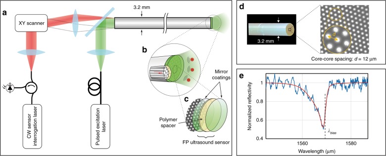
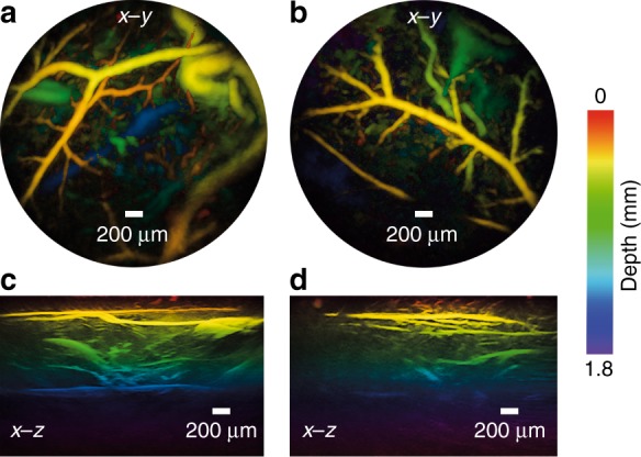
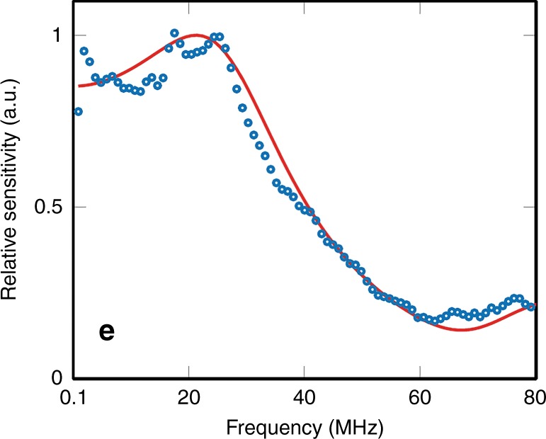
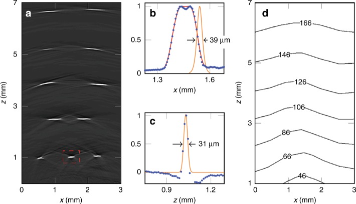
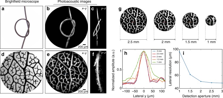
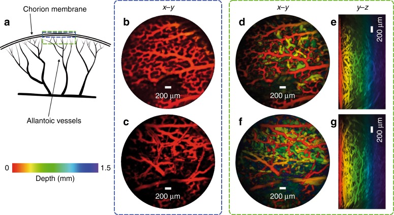
A miniature forward-viewing endoscopic probe that provides high-resolution 3D photoacoustic images is demonstrated. The probe is of outer diameter 3.2 mm and comprised of a transparent Fabry-Pérot (FP) polymer-film ultrasound sensor that is located at the distal end of a rigid optical fiber bundle. Excitation laser pulses are coupled simultaneously into all cores of the bundle and are transmitted through the FP sensor to provide wide-field tissue illumination at the distal end. The resulting photoacoustic waves are mapped in 2D by sequentially scanning the input end of the bundle with an interrogation laser beam in order to individually address different points on the FP sensor. In this way, the sensor acts as a high-density ultrasound array that is comprised of 50,000 individual elements, each of which is 12 µm in diameter, within the 3.2 mm diameter footprint of the probe. The fine spatial sampling that this affords, along with the wide bandwidth ( 34 MHz) of the sensor, enables a high-resolution photoacoustic image to be reconstructed. The measured on-axis lateral resolution of the probe was depth-dependent and ranged from 45-170 µm for depths between 1 and 7 mm, and the vertical resolution was 31 µm over the same depth range. The system was evaluated by acquiring 3D images of absorbing phantoms and the microvascular anatomies of a duck embryo and mouse skin. Excellent image fidelity was demonstrated. It is anticipated that this type of probe could find application as a tool for guiding laparoscopic procedures, fetal surgery and other minimally invasive interventions that require a millimeter-scale forward-viewing 3D photoacoustic imaging probe.
展示了一种能提供高分辨率三维光声图像的微型前视内窥镜探头。该探头外径为3.2毫米,由一个透明的法布里-珀罗(FP)聚合物薄膜超声传感器组成,该传感器位于刚性光纤束的远端。激发激光脉冲同时耦合到光纤束的所有纤芯中,并通过FP传感器传输,以在远端提供宽场组织照明。通过用探测激光束顺序扫描光纤束的输入端,从而单独寻址FP传感器上的不同点,将产生的光声波映射成二维图像。通过这种方式,该传感器充当了一个高密度超声阵列,在探头直径3.2毫米的范围内由50000个单独的元件组成,每个元件的直径为12微米。这种精细的空间采样,连同传感器的宽带宽(34兆赫),使得能够重建高分辨率的光声图像。探头在轴向上测得的横向分辨率与深度有关,在1至7毫米的深度范围内,分辨率范围为45至170微米,在相同深度范围内垂直分辨率为31微米。通过获取吸收性体模以及鸭胚胎和小鼠皮肤的微血管解剖结构的三维图像对该系统进行了评估。结果表明图像保真度极佳。预计这种类型的探头可作为一种工具,用于指导腹腔镜手术、胎儿手术以及其他需要毫米级前视三维光声成像探头的微创干预。