National Cancer Center/National Clinical Research Center for Cancer/Cancer Hospital, Chinese Academy of Medical Sciences and Peking Union Medical College, Beijing, 100021, China.
Med Phys. 2019 Jan;46(1):56-64. doi: 10.1002/mp.13262. Epub 2018 Nov 23.
To develop a method for predicting optimal dose distributions, given the planning image and segmented anatomy, by applying deep learning techniques to a database of previously optimized and approved Intensity-modulated radiation therapy treatment plans.
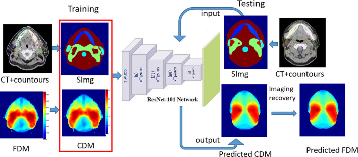
Eighty cases of early-stage nasopharyngeal cancer (NPC) were included in the study. Seventy cases were chosen randomly as the training set and the remaining as the test set. The inputs were the images with structures, with each target and organs at risk (OARs) assigned a unique label. The outputs were dose maps, including coarse dose maps and converted fine dose maps (FDM) from convolution. Two types of input images with structures were used in the model building. One type of input included the images (with associated structures) without manipulation. The second type of input involved modifying the image gray label with information from radiation beam geometry. ResNet101 was chosen as the deep learning network for both. The accuracy of predicted dose distributions was evaluated against the corresponding dose as used in the clinic. A global three-dimensional gamma analysis was calculated for the evaluation.
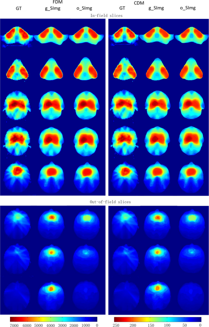
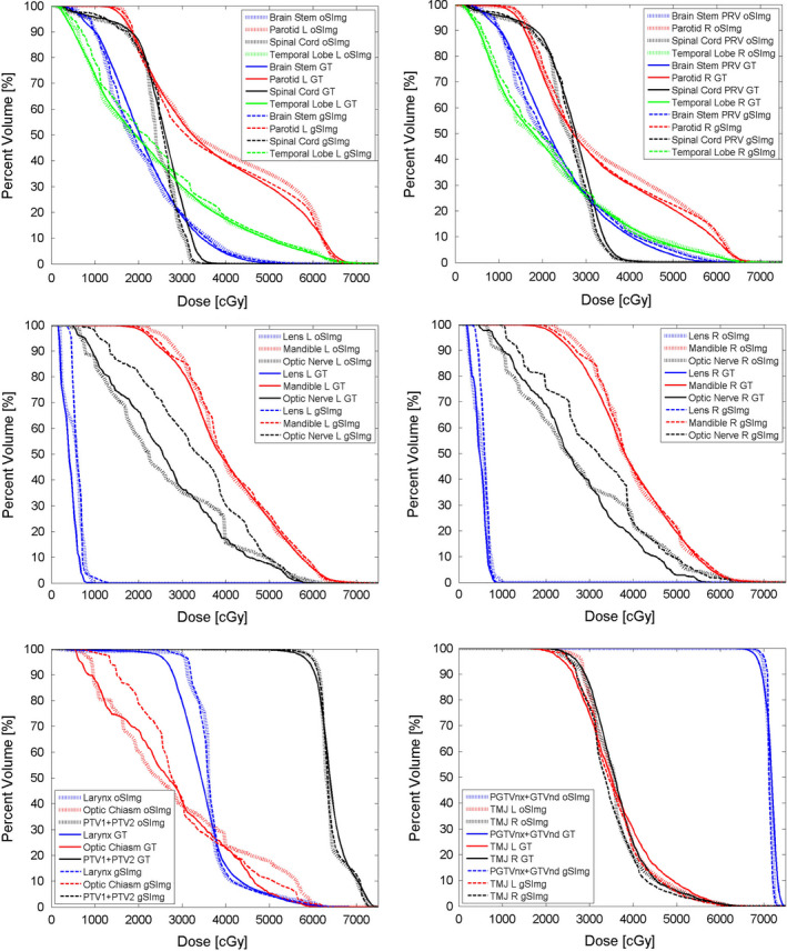
The proposed model trained with the two different sets of input images and structures could both predict patient-specific dose distributions accurately. For the out-of-field dose distributions, the model obtained from the input with radiation geometry performed better (dose difference in %, 4.7 ± 6.1% vs 5.5 ± 7.9%, P < 0.05). The mean Gamma pass rates of dose distributions predicted with both types of input were comparable for most OARs (P > 0.05), except for the bilateral optic nerves and the optic chiasm.
The proposed system with radiation geometry added to the input is a promising method to generate patient-specific dose distributions for radiotherapy. It can be applied to obtain the dose distributions slice-by-slice for planning quality assurance and for guiding automated planning.
通过将深度学习技术应用于先前优化和批准的调强放射治疗计划数据库,开发一种从计划图像和分割解剖结构中预测最佳剂量分布的方法。
本研究纳入 80 例早期鼻咽癌(NPC)患者。70 例随机选择作为训练集,其余作为测试集。输入是带有结构的图像,每个目标和危及器官(OARs)都被赋予一个唯一的标签。输出是剂量图,包括卷积生成的粗略剂量图和转换后的精细剂量图(FDM)。在模型构建中使用了两种结构的输入图像。一种输入类型包括未经过处理的图像(带有相关结构)。第二种输入类型涉及使用放射束几何信息修改图像灰度标签。两种输入图像都选择 ResNet101 作为深度学习网络。通过与临床使用的剂量进行比较来评估预测剂量分布的准确性。计算了全局三维伽马分析进行评估。
使用两种不同的输入图像和结构集训练的提出的模型都可以准确预测患者特定的剂量分布。对于场外剂量分布,从具有放射几何形状的输入中获得的模型表现更好(剂量差异百分比,4.7±6.1%比 5.5±7.9%,P<0.05)。两种输入类型预测的剂量分布的平均伽马通过率在大多数 OARs 中相当(P>0.05),除了双侧视神经和视交叉。
将放射几何形状添加到输入中的所提出的系统是生成放射治疗患者特定剂量分布的有前途的方法。它可用于为计划质量保证和指导自动规划获得逐片剂量分布。