Chair of Biomedical Physics, Department of Physics and Munich School of BioEngineering, Technical University of Munich, 85748, Garching, Germany.
Department of Diagnostic and Interventional Radiology, Klinikum rechts der Isar, Technical University of Munich, 81675, München, Germany.
Sci Rep. 2018 Nov 6;8(1):16394. doi: 10.1038/s41598-018-34809-6.
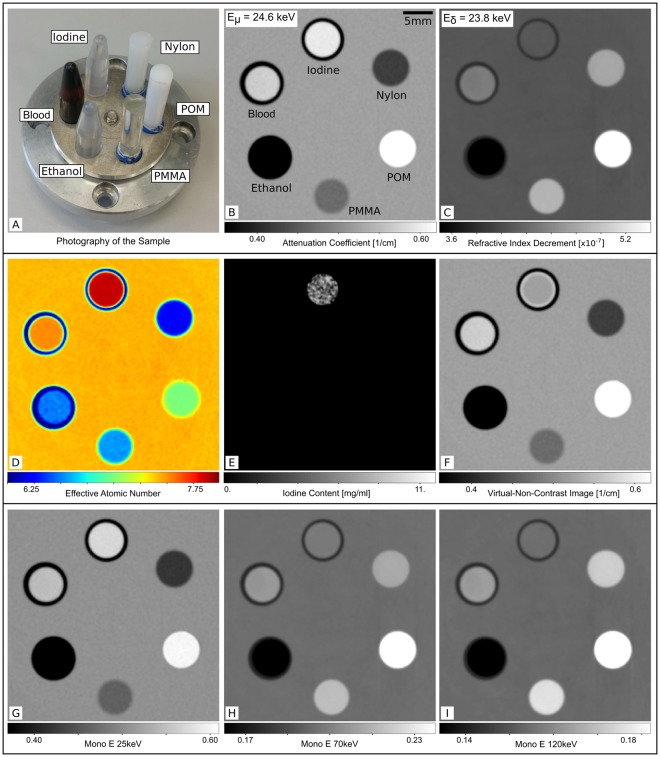
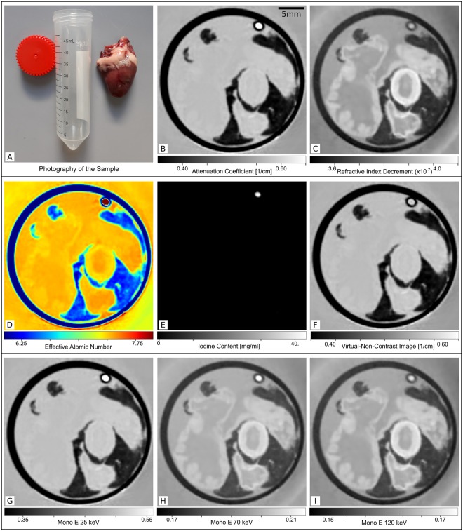
Dual-energy CT has opened up a new level of quantitative X-ray imaging for many diagnostic applications. The energy dependence of the X-ray attenuation is the key to quantitative material decomposition of the volume under investigation. This material decomposition allows the calculation of virtual native images in contrast enhanced angiography, virtual monoenergetic images for beam-hardening artifact reduction and quantitative material maps, among others. These visualizations have been proven beneficial for various diagnostic questions. Here, we demonstrate a new method of 'virtual dual-energy CT' employing grating-based phase-contrast for quantitative material decomposition. Analogue to the measurement at two different energies, the applied phase-contrast measurement approach yields dual information in form of a phase-shift and an attenuation image. Based on these two image channels, all known dual-energy applications can be demonstrated with our technique. While still in a preclinical state, the method features the important advantages of direct access to the electron density via the phase image, simultaneous availability of the conventional attenuation image at the full energy spectrum and therefore inherently registered image channels. The transfer of this signal extraction approach to phase-contrast data multiplies the diagnostic information gained within a single CT acquisition. The method is demonstrated with a phantom consisting of exemplary solid and fluid materials as well as a chicken heart with an iodine filled tube simulating a vessel. For this first demonstration all measurements have been conducted at a compact laser-undulator synchrotron X-ray source with a tunable X-ray energy and a narrow spectral bandwidth, to validate the quantitativeness of the processing approach.
双能 CT 为许多诊断应用开辟了新的定量 X 射线成像水平。X 射线衰减的能量依赖性是对所研究体积进行定量物质分解的关键。这种物质分解允许计算对比度增强血管造影中的虚拟原生图像、用于束硬化伪影减少的虚拟单能量图像以及定量物质图等。这些可视化效果已被证明对各种诊断问题有益。在这里,我们展示了一种使用基于光栅的相衬进行定量物质分解的“虚拟双能 CT”新方法。类似于在两种不同能量下的测量,所应用的相衬测量方法以相移和衰减图像的形式提供双重信息。基于这两个图像通道,我们的技术可以展示所有已知的双能应用。虽然仍处于临床前阶段,但该方法具有通过相位图像直接访问电子密度、同时在全能谱上获得常规衰减图像以及因此固有注册图像通道的重要优势。将这种信号提取方法应用于相衬数据可在单次 CT 采集内增加获得的诊断信息量。该方法使用由示例固体和流体材料以及充满碘的管模拟血管的鸡心组成的幻影进行了演示。对于这个首次演示,所有测量都是在具有可调谐 X 射线能量和窄光谱带宽的紧凑型激光-直线加速器同步加速器 X 射线源上进行的,以验证处理方法的定量性。