• 文献检索
  • 文档翻译
  • 深度研究
  • 学术资讯
  • Suppr Zotero 插件Zotero 插件
  • 邀请有礼
  • 套餐&价格
  • 历史记录
应用&插件
Suppr Zotero 插件Zotero 插件浏览器插件Mac 客户端Windows 客户端微信小程序
定价
高级版会员购买积分包购买API积分包
服务
文献检索文档翻译深度研究API 文档MCP 服务
关于我们
关于 Suppr公司介绍联系我们用户协议隐私条款
关注我们

Suppr 超能文献

核心技术专利:CN118964589B侵权必究
粤ICP备2023148730 号-1Suppr @ 2026

文献检索

告别复杂PubMed语法,用中文像聊天一样搜索,搜遍4000万医学文献。AI智能推荐,让科研检索更轻松。

立即免费搜索

文件翻译

保留排版,准确专业,支持PDF/Word/PPT等文件格式,支持 12+语言互译。

免费翻译文档

深度研究

AI帮你快速写综述,25分钟生成高质量综述,智能提取关键信息,辅助科研写作。

立即免费体验

基于 X 射线计算机断层扫描的成年硬骨鱼类定量形态测量分析。

Quantitative morphometric analysis of adult teleost fish by X-ray computed tomography.

机构信息

Institute for Photon Science and Synchrotron Radiation, Karlsruhe Institute of Technology, Hermann-von-Helmholtz-Platz 1, Eggenstein-Leopoldshafen, Germany.

Centre for Organismal Studies, COS, Heidelberg University, Im Neunheimer Feld 230, Heidelberg, Germany.

出版信息

Sci Rep. 2018 Nov 8;8(1):16531. doi: 10.1038/s41598-018-34848-z.

DOI:10.1038/s41598-018-34848-z
PMID:30410001
原文链接:https://pmc.ncbi.nlm.nih.gov/articles/PMC6224569/
Abstract

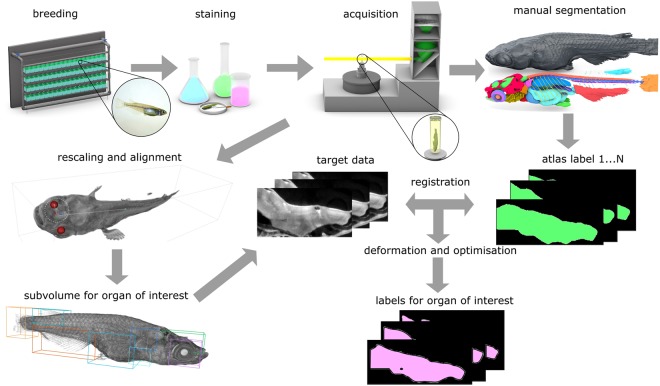
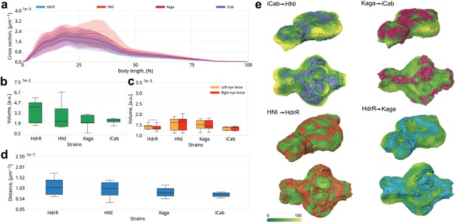
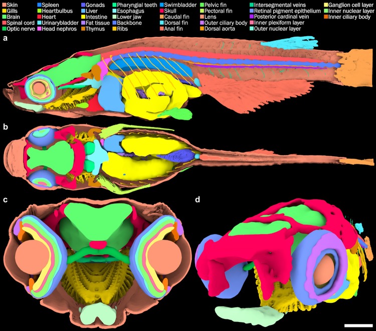
Vertebrate models provide indispensable paradigms to study development and disease. Their analysis requires a quantitative morphometric study of the body, organs and tissues. This is often impeded by pigmentation and sample size. X-ray micro-computed tomography (micro-CT) allows high-resolution volumetric tissue analysis, largely independent of sample size and transparency to visual light. Importantly, micro-CT data are inherently quantitative. We report a complete pipeline of high-throughput 3D data acquisition and image analysis, including tissue preparation and contrast enhancement for micro-CT imaging down to cellular resolution, automated data processing and organ or tissue segmentation that is applicable to comparative 3D morphometrics of small vertebrates. Applied to medaka fish, we first create an annotated anatomical atlas of the entire body, including inner organs as a quantitative morphological description of an adult individual. This atlas serves as a reference model for comparative studies. Using isogenic medaka strains we show that comparative 3D morphometrics of individuals permits identification of quantitative strain-specific traits. Thus, our pipeline enables high resolution morphological analysis as a basis for genotype-phenotype association studies of complex genetic traits in vertebrates.

摘要

脊椎动物模型为研究发育和疾病提供了不可或缺的范例。对它们的分析需要对身体、器官和组织进行定量形态计量学研究。这通常受到色素沉着和样本大小的限制。X 射线微计算机断层扫描(micro-CT)允许对组织进行高分辨率的体积分析,在很大程度上不受样本大小和可见光透明度的影响。重要的是,micro-CT 数据本质上是定量的。我们报告了一个完整的高通量 3D 数据采集和图像分析流水线,包括组织准备和对比增强,以实现高达细胞分辨率的 micro-CT 成像、自动数据处理以及适用于小型脊椎动物比较 3D 形态计量学的器官或组织分割。我们将其应用于斑马鱼,首先创建了整个身体的注释解剖图谱,包括内部器官,作为成年个体的定量形态描述。该图谱作为比较研究的参考模型。使用同基因的斑马鱼品系,我们表明个体的比较 3D 形态计量学可以识别定量的品系特异性特征。因此,我们的流水线能够进行高分辨率形态分析,作为脊椎动物复杂遗传特征的基因型-表型关联研究的基础。

https://cdn.ncbi.nlm.nih.gov/pmc/blobs/3f81/6224569/77b1aabd5b73/41598_2018_34848_Fig5_HTML.jpg
https://cdn.ncbi.nlm.nih.gov/pmc/blobs/3f81/6224569/555d21abc16e/41598_2018_34848_Fig1_HTML.jpg
https://cdn.ncbi.nlm.nih.gov/pmc/blobs/3f81/6224569/3f7fac1426a3/41598_2018_34848_Fig2_HTML.jpg
https://cdn.ncbi.nlm.nih.gov/pmc/blobs/3f81/6224569/3fdc3f470091/41598_2018_34848_Fig3_HTML.jpg
https://cdn.ncbi.nlm.nih.gov/pmc/blobs/3f81/6224569/7f55561f8fe5/41598_2018_34848_Fig4_HTML.jpg
https://cdn.ncbi.nlm.nih.gov/pmc/blobs/3f81/6224569/77b1aabd5b73/41598_2018_34848_Fig5_HTML.jpg

相似文献

1
Quantitative morphometric analysis of adult teleost fish by X-ray computed tomography.基于 X 射线计算机断层扫描的成年硬骨鱼类定量形态测量分析。
Sci Rep. 2018 Nov 8;8(1):16531. doi: 10.1038/s41598-018-34848-z.
2
A method of 2D/3D registration of a statistical mouse atlas with a planar X-ray projection and an optical photo.一种将统计鼠标图谱的二维/三维图像与平面 X 射线投影和光学照片进行配准的方法。
Med Image Anal. 2013 May;17(4):401-16. doi: 10.1016/j.media.2013.02.009. Epub 2013 Mar 5.
3
Technical note: Three-dimensional imaging of rumen tissue for morphometric analysis using micro-computed tomography.技术说明:使用微型计算机断层扫描对瘤胃组织进行形态计量分析的三维成像
J Dairy Sci. 2014 Dec;97(12):7691-6. doi: 10.3168/jds.2014-8374. Epub 2014 Sep 26.
4
3D Atlas of the Pituitary Gland of the Model Fish Medaka ().模式鱼类青鳉垂体的 3D 图谱 ()。
Front Endocrinol (Lausanne). 2021 Aug 23;12:719843. doi: 10.3389/fendo.2021.719843. eCollection 2021.
5
Structural analysis of the female reptile reproductive system by micro-computed tomography and optical coherence tomography†.采用微计算机断层扫描和光学相干断层扫描技术对雌性爬行动物生殖系统的结构分析。
Biol Reprod. 2024 Jun 12;110(6):1077-1085. doi: 10.1093/biolre/ioae039.
6
Femur statistical atlas construction based on two-level 3D non-rigid registration.基于两级三维非刚性配准的股骨统计图谱构建
Comput Aided Surg. 2009;14(4-6):83-99. doi: 10.3109/10929080903246543.
7
A novel 3D mouse embryo atlas based on micro-CT.基于微 CT 的新型 3D 鼠胚胎图谱
Development. 2012 Sep;139(17):3248-56. doi: 10.1242/dev.082016.
8
Improved middle-ear soft-tissue visualization using synchrotron radiation phase-contrast imaging.使用同步辐射相衬成像改善中耳软组织可视化。
Hear Res. 2017 Oct;354:1-8. doi: 10.1016/j.heares.2017.08.001. Epub 2017 Aug 5.
9
Potential and limitations of X-Ray micro-computed tomography in arthropod neuroanatomy: a methodological and comparative survey.X射线显微计算机断层扫描在节肢动物神经解剖学中的潜力与局限:一项方法学与比较性研究
J Comp Neurol. 2015 Jun 1;523(8):1281-95. doi: 10.1002/cne.23741. Epub 2015 Mar 2.
10
Constructing and optimizing 3D atlases from 2D data with application to the developing mouse brain.从二维数据构建和优化 3D 图谱及其在发育中老鼠大脑中的应用。
Elife. 2021 Feb 11;10:e61408. doi: 10.7554/eLife.61408.

引用本文的文献

1
The sex of organ geometry.器官几何形状的性别。
Nature. 2024 Jun;630(8016):392-400. doi: 10.1038/s41586-024-07463-4. Epub 2024 May 29.
2
Visualisation of gene expression within the context of tissues using an X-ray computed tomography-based multimodal approach.基于 X 射线计算机断层扫描的多模态方法在组织中可视化基因表达。
Sci Rep. 2024 Apr 12;14(1):8543. doi: 10.1038/s41598-024-58766-5.
3
Enhanced contrast synchrotron X-ray microtomography for describing skeleton-associated soft tissue defects in zebrafish mutants.增强对比同步辐射 X 射线微断层扫描用于描述斑马鱼突变体中与骨骼相关的软组织缺陷。

本文引用的文献

1
Development of caudal structures of a morphogenetic mutant (Da) in the teleost fish, medaka (Oryzias latipes).硬骨鱼青鳉(Oryzias latipes)形态发生突变体(Da)尾部结构的发育
J Morphol. 1990 Aug;205(2):219-232. doi: 10.1002/jmor.1052050209.
2
MicroCT-based phenomics in the zebrafish skeleton reveals virtues of deep phenotyping in a distributed organ system.基于微计算机断层扫描的斑马鱼骨骼表型组学研究揭示了分布式器官系统中深度表型分析的优点。
Elife. 2017 Sep 8;6:e26014. doi: 10.7554/eLife.26014.
3
Automatic segmentation and quantification of the cardiac structures from non-contrast-enhanced cardiac CT scans.
Front Endocrinol (Lausanne). 2023 Mar 6;14:1108916. doi: 10.3389/fendo.2023.1108916. eCollection 2023.
4
A wide-field micro-computed tomography detector: micron resolution at half-centimetre scale.宽视场微计算机断层扫描探测器:半厘米尺度下的微米分辨率。
J Synchrotron Radiat. 2022 Mar 1;29(Pt 2):505-514. doi: 10.1107/S160057752101287X. Epub 2022 Jan 19.
5
Turning the Curve Into Straight: Phenogenetics of the Spine Morphology and Coordinate Maintenance in the Zebrafish.化曲为直:斑马鱼脊柱形态与坐标维持的表型遗传学
Front Cell Dev Biol. 2022 Jan 26;9:801652. doi: 10.3389/fcell.2021.801652. eCollection 2021.
6
Integration of multiple imaging platforms to uncover cardiovascular defects in adult zebrafish.整合多种成像平台以揭示成年斑马鱼的心血管缺陷。
Cardiovasc Res. 2022 Sep 20;118(12):2665-2687. doi: 10.1093/cvr/cvab310.
7
Normal stages of embryonic development of a brood parasite, the rosy bitterling Rhodeus ocellatus (Teleostei: Cypriniformes).正常的寄生性鱼类胚胎发育阶段,红鳍鲫(鲤形目:鲤科)。
J Morphol. 2021 Jun;282(6):783-819. doi: 10.1002/jmor.21335. Epub 2021 Apr 2.
8
Introducing Biomedisa as an open-source online platform for biomedical image segmentation.介绍 Biomedisa,这是一个用于生物医学图像分割的开源在线平台。
Nat Commun. 2020 Nov 4;11(1):5577. doi: 10.1038/s41467-020-19303-w.
9
Synchrotron microtomography applied to the volumetric analysis of internal structures of Thoropa miliaris tadpoles.同步辐射微断层扫描技术在分析密点麻蜥蝌蚪内部结构体积中的应用。
Sci Rep. 2020 Nov 3;10(1):18934. doi: 10.1038/s41598-020-75993-8.
10
Whole Animal Imaging of Drosophila melanogaster using Microcomputed Tomography.使用微型计算机断层扫描对黑腹果蝇进行全动物成像。
J Vis Exp. 2020 Sep 2(163). doi: 10.3791/61515.
从非增强心脏CT扫描中自动分割和量化心脏结构。
Phys Med Biol. 2017 May 7;62(9):3798-3813. doi: 10.1088/1361-6560/aa63cb. Epub 2017 Mar 1.
4
A Novel Mouse Segmentation Method Based on Dynamic Contrast Enhanced Micro-CT Images.一种基于动态对比增强微型计算机断层扫描图像的新型小鼠分割方法。
PLoS One. 2017 Jan 6;12(1):e0169424. doi: 10.1371/journal.pone.0169424. eCollection 2017.
5
Three-dimensional microCT imaging of mouse development from early post-implantation to early postnatal stages.从小鼠植入后早期到出生后早期发育阶段的三维显微CT成像。
Dev Biol. 2016 Nov 15;419(2):229-236. doi: 10.1016/j.ydbio.2016.09.011. Epub 2016 Sep 23.
6
High-throughput discovery of novel developmental phenotypes.新型发育表型的高通量发现
Nature. 2016 Sep 22;537(7621):508-514. doi: 10.1038/nature19356. Epub 2016 Sep 14.
7
Optimal Contrast Agent Staining of Ligaments and Tendons for X-Ray Computed Tomography.用于X射线计算机断层扫描的韧带和肌腱的最佳造影剂染色
PLoS One. 2016 Apr 14;11(4):e0153552. doi: 10.1371/journal.pone.0153552. eCollection 2016.
8
Contrast-Enhanced X-Ray Micro-Computed Tomography as a Versatile Method for Anatomical Studies of Adult Zebrafish.对比增强X射线显微计算机断层扫描作为成年斑马鱼解剖学研究的通用方法
Zebrafish. 2016 Aug;13(4):310-6. doi: 10.1089/zeb.2016.1245. Epub 2016 Apr 8.
9
Grating-based X-ray Dark-field Computed Tomography of Living Mice.基于光栅的活体小鼠X射线暗场计算机断层扫描
EBioMedicine. 2015 Aug 13;2(10):1500-6. doi: 10.1016/j.ebiom.2015.08.014. eCollection 2015 Oct.
10
Micro-computed tomography (μCT) as a novel method in ecotoxicology--determination of morphometric and somatic data in rainbow trout (Oncorhynchus mykiss).微计算机断层扫描(μCT)作为一种新的生态毒理学方法——虹鳟鱼(Oncorhynchus mykiss)形态计量和体数据的测定。
Sci Total Environ. 2016 Feb 1;543(Pt A):135-139. doi: 10.1016/j.scitotenv.2015.11.020. Epub 2015 Nov 12.