Jung HaeWon, Liu Tao, Liu Jianfei, Huryn Laryssa A, Tam Johnny
National Eye Institute, National Institutes of Health, Bethesda, MD, 20892, USA.
Commun Biol. 2018 Nov 14;1:189. doi: 10.1038/s42003-018-0190-8. eCollection 2018.
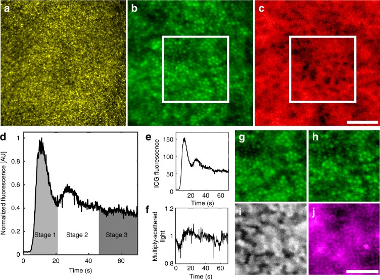
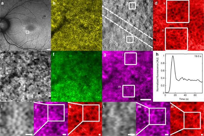
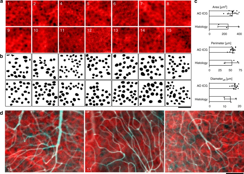
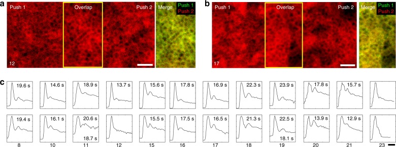
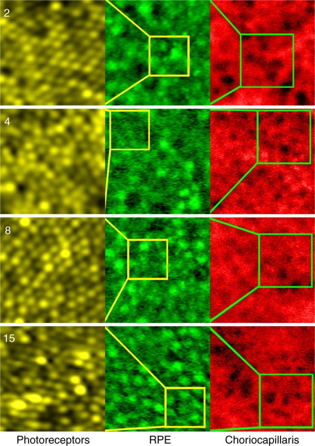
Visualizing the cellular manifestation of disease has recently been aided by an increasing number of adaptive optics (AO)-based imaging modalities developed for the living human eye. However, simultaneous visualization of multiple, interacting cell types within a complete neural-epithelial-vascular complex has proven challenging. By incorporating AO with indocyanine green angiography, we demonstrate the possibility of imaging photoreceptors, retinal pigment epithelial cells, and choriocapillaris in the living human eye. Unexpectedly, we found that there was uptake of indocyanine green dye into the retinal pigment epithelial cells in the earliest phases of imaging, which formed the basis for devising a strategy to visualize the choriocapillaris. Our results expand the range of applications for an existing, FDA-approved, systemically injected fluorescent dye. The combined multimodal approach can be used to evaluate the complete outer retinal complex at the cellular level, a transformative step toward revealing the in vivo cellular status of neurodegenerative conditions and blinding diseases.
近年来,为活体人眼开发的越来越多基于自适应光学(AO)的成像方式有助于可视化疾病的细胞表现。然而,在完整的神经 - 上皮 - 血管复合体中同时可视化多种相互作用的细胞类型已被证明具有挑战性。通过将AO与吲哚菁绿血管造影相结合,我们证明了在活体人眼中对光感受器、视网膜色素上皮细胞和脉络膜毛细血管进行成像的可能性。出乎意料的是,我们发现在成像的最早阶段吲哚菁绿染料被视网膜色素上皮细胞摄取,这为设计一种可视化脉络膜毛细血管的策略奠定了基础。我们的结果扩展了一种现有的、经美国食品药品监督管理局(FDA)批准的全身注射荧光染料的应用范围。这种联合多模态方法可用于在细胞水平评估完整的视网膜外层复合体,这是朝着揭示神经退行性疾病和致盲疾病的体内细胞状态迈出的变革性一步。