Liu Jianfei, Jung HaeWon, Dubra Alfredo, Tam Johnny
Ophthalmic Genetics and Visual Function Branch, National Eye Institute, National Institutes of Health, Bethesda, Maryland, United States.
Department of Ophthalmology, Stanford University, Palo Alto, California, United States.
Invest Ophthalmol Vis Sci. 2017 Sep 1;58(11):4477-4489. doi: 10.1167/iovs.16-21003.
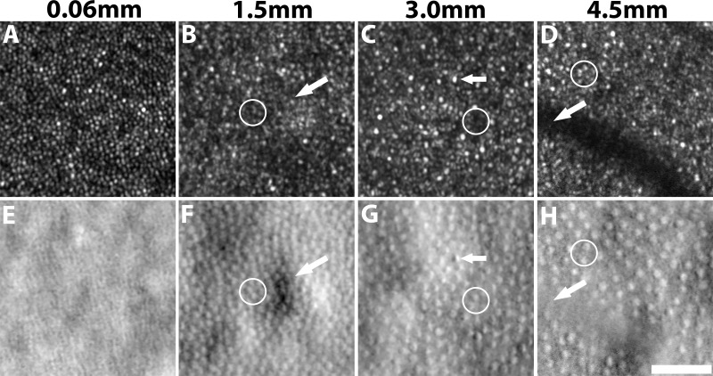
Adaptive optics scanning light ophthalmoscopy (AOSLO) has enabled quantification of the photoreceptor mosaic in the living human eye using metrics such as cell density and average spacing. These rely on the identification of individual cells. Here, we demonstrate a novel approach for computer-aided identification of cone photoreceptors on nonconfocal split detection AOSLO images.
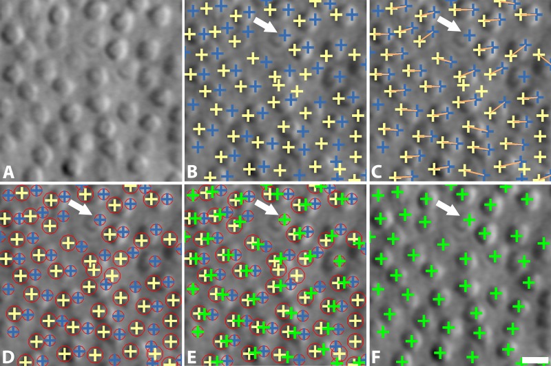
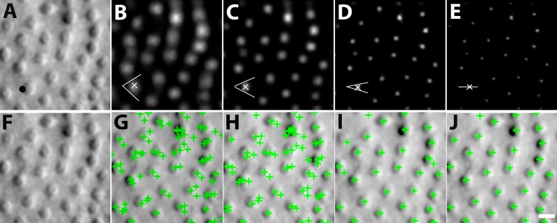
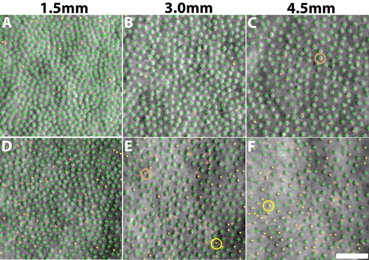
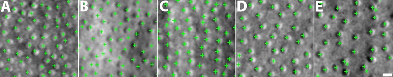
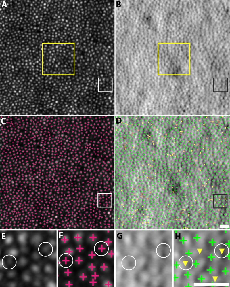
Algorithms for identification of cone photoreceptors were developed, based on multiscale circular voting (MSCV) in combination with a priori knowledge that split detection images resemble Nomarski differential interference contrast images, in which dark and bright regions are present on the two sides of each cell. The proposed algorithm locates dark and bright region pairs, iteratively refining the identification across multiple scales. Identification accuracy was assessed in data from 10 subjects by comparing automated identifications with manual labeling, followed by computation of density and spacing metrics for comparison to histology and published data.
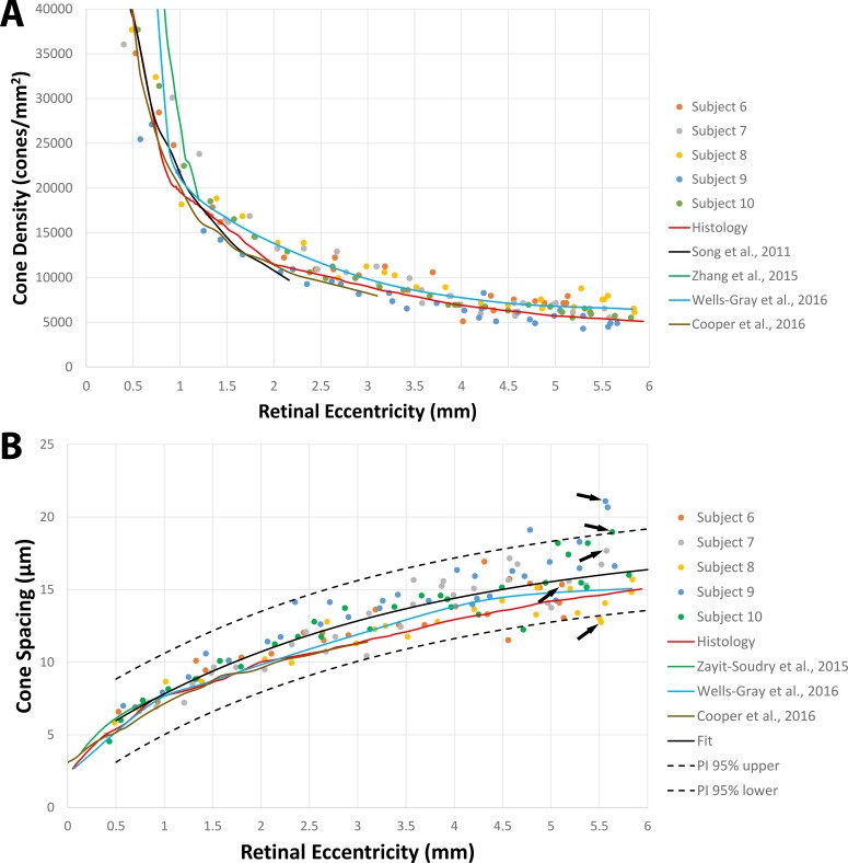
There was good agreement between manual and automated cone identifications with overall recall, precision, and F1 score of 92.9%, 90.8%, and 91.8%, respectively. On average, computed density and spacing values using automated identification were within 10.7% and 11.2% of the expected histology values across eccentricities ranging from 0.5 to 6.2 mm. There was no statistically significant difference between MSCV-based and histology-based density measurements (P = 0.96, Kolmogorov-Smirnov 2-sample test).
MSCV can accurately detect cone photoreceptors on split detection images across a range of eccentricities, enabling quick, objective estimation of photoreceptor mosaic metrics, which will be important for future clinical trials utilizing adaptive optics.
自适应光学扫描光检眼镜(AOSLO)已能够使用细胞密度和平均间距等指标对活体人眼的光感受器镶嵌结构进行量化。这些指标依赖于对单个细胞的识别。在此,我们展示了一种在非共焦分裂检测AOSLO图像上进行计算机辅助视锥光感受器识别的新方法。
基于多尺度循环投票(MSCV)并结合先验知识开发了视锥光感受器识别算法,该先验知识是分裂检测图像类似于诺马斯基微分干涉对比图像,其中每个细胞的两侧存在暗区和亮区。所提出的算法定位暗区和亮区对,在多个尺度上迭代优化识别。通过将自动识别与手动标记进行比较,评估了10名受试者数据中的识别准确性,随后计算密度和间距指标以与组织学和已发表数据进行比较。
手动和自动视锥识别之间具有良好的一致性,总体召回率、精确率和F1分数分别为92.9%、90.8%和91.8%。平均而言,使用自动识别计算的密度和间距值在偏心度范围为0.5至6.2毫米时,与预期组织学值的偏差在10.7%和11.2%以内。基于MSCV的密度测量与基于组织学的密度测量之间无统计学显著差异(P = 0.96,柯尔莫哥洛夫 - 斯米尔诺夫双样本检验)。
MSCV能够在一系列偏心度的分裂检测图像上准确检测视锥光感受器,从而能够快速、客观地估计光感受器镶嵌结构指标,这对于未来利用自适应光学的临床试验将具有重要意义。