Kazuhiro Koshino, Werner Rudolf A, Toriumi Fujio, Javadi Mehrbod S, Pomper Martin G, Solnes Lilja B, Verde Franco, Higuchi Takahiro, Rowe Steven P
Department of Biomedical Imaging, National Cardiovascular and Cerebral Research Center, Suita, Japan.
The Russell H. Morgan Department of Radiology and Radiological Science, Division of Nuclear Medicine and Molecular Imaging, Johns Hopkins School University of Medicine, Baltimore, MD.
Tomography. 2018 Dec;4(4):159-163. doi: 10.18383/j.tom.2018.00042.
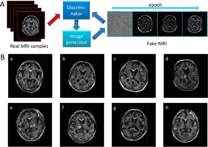
Even as medical data sets become more publicly accessible, most are restricted to specific medical conditions. Thus, data collection for machine learning approaches remains challenging, and synthetic data augmentation, such as generative adversarial networks (GAN), may overcome this hurdle. In the present quality control study, deep convolutional GAN (DCGAN)-based human brain magnetic resonance (MR) images were validated by blinded radiologists. In total, 96 T1-weighted brain images from 30 healthy individuals and 33 patients with cerebrovascular accident were included. A training data set was generated from the T1-weighted images and DCGAN was applied to generate additional artificial brain images. The likelihood that images were DCGAN-created versus acquired was evaluated by 5 radiologists (2 neuroradiologists [NRs], vs 3 non-neuroradiologists [NNRs]) in a binary fashion to identify real vs created images. Images were selected randomly from the data set (variation of created images, 40%-60%). None of the investigated images was rated as unknown. Of the created images, the NRs rated 45% and 71% as real magnetic resonance imaging images (NNRs, 24%, 40%, and 44%). In contradistinction, 44% and 70% of the real images were rated as generated images by NRs (NNRs, 10%, 17%, and 27%). The accuracy for the NRs was 0.55 and 0.30 (NNRs, 0.83, 0.72, and 0.64). DCGAN-created brain MR images are similar enough to acquired MR images so as to be indistinguishable in some cases. Such an artificial intelligence algorithm may contribute to synthetic data augmentation for "data-hungry" technologies, such as supervised machine learning approaches, in various clinical applications.
尽管医学数据集越来越容易公开获取,但大多数数据集都局限于特定的医疗状况。因此,机器学习方法的数据收集仍然具有挑战性,而合成数据增强技术,如生成对抗网络(GAN),可能会克服这一障碍。在本质量控制研究中,基于深度卷积GAN(DCGAN)生成的人脑磁共振(MR)图像由不知情的放射科医生进行验证。总共纳入了来自30名健康个体和33名脑血管意外患者的96张T1加权脑图像。从T1加权图像生成训练数据集,并应用DCGAN生成额外的人工脑图像。5名放射科医生(2名神经放射科医生[NRs]和3名非神经放射科医生[NNRs])以二元方式评估图像是由DCGAN生成还是实际采集的,以识别真实图像与生成图像。从数据集中随机选择图像(生成图像的变化范围为40%-60%)。所有被调查的图像均未被评为无法确定。在生成的图像中,NRs将45%和71%评为真实的磁共振成像图像(NNRs分别为24%、40%和44%)。相反,NRs将44%和70%的真实图像评为生成图像(NNRs分别为10%、17%和27%)。NRs的准确率为0.55和0.30(NNRs分别为0.83、0.72和0.64)。DCGAN生成的脑MR图像与采集的MR图像非常相似,以至于在某些情况下难以区分。这种人工智能算法可能有助于为各种临床应用中的“数据饥渴型”技术(如监督机器学习方法)进行合成数据增强。