• 文献检索
  • 文档翻译
  • 深度研究
  • 学术资讯
  • Suppr Zotero 插件Zotero 插件
  • 邀请有礼
  • 套餐&价格
  • 历史记录
应用&插件
Suppr Zotero 插件Zotero 插件浏览器插件Mac 客户端Windows 客户端微信小程序
定价
高级版会员购买积分包购买API积分包
服务
文献检索文档翻译深度研究API 文档MCP 服务
关于我们
关于 Suppr公司介绍联系我们用户协议隐私条款
关注我们

Suppr 超能文献

核心技术专利:CN118964589B侵权必究
粤ICP备2023148730 号-1Suppr @ 2026

文献检索

告别复杂PubMed语法,用中文像聊天一样搜索,搜遍4000万医学文献。AI智能推荐,让科研检索更轻松。

立即免费搜索

文件翻译

保留排版,准确专业,支持PDF/Word/PPT等文件格式,支持 12+语言互译。

免费翻译文档

深度研究

AI帮你快速写综述,25分钟生成高质量综述,智能提取关键信息,辅助科研写作。

立即免费体验

计算机辅助激光解剖:一种利用图像分析工具的显微解剖工作流程。

Computer-Aided Laser Dissection: A Microdissection Workflow Leveraging Image Analysis Tools.

作者信息

Hipp Jason D, Johann Donald J, Chen Yun, Madabhushi Anant, Monaco James, Cheng Jerome, Rodriguez-Canales Jaime, Stumpe Martin C, Riedlinger Greg, Rosenberg Avi Z, Hanson Jeffrey C, Kunju Lakshmi P, Emmert-Buck Michael R, Balis Ulysses J, Tangrea Michael A

机构信息

Laboratory of Pathology, National Cancer Institute, Bethesda, MD, USA.

Google Inc., Mountain View, CA, USA.

出版信息

J Pathol Inform. 2018 Dec 11;9:45. doi: 10.4103/jpi.jpi_60_18. eCollection 2018.

DOI:10.4103/jpi.jpi_60_18
PMID:30622835
原文链接:https://pmc.ncbi.nlm.nih.gov/articles/PMC6298131/
Abstract

INTRODUCTION

The development and application of new molecular diagnostic assays based on next-generation sequencing and proteomics require improved methodologies for procurement of target cells from histological sections. Laser microdissection can successfully isolate distinct cells from tissue specimens based on visual selection for many research and clinical applications. However, this can be a daunting task when a large number of cells are required for molecular analysis or when a sizeable number of specimens need to be evaluated.

MATERIALS AND METHODS

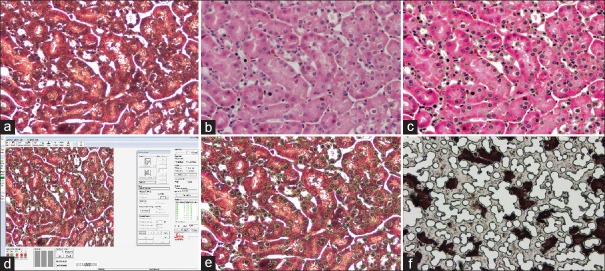
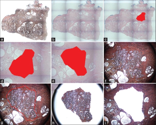
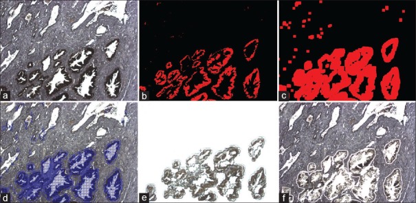
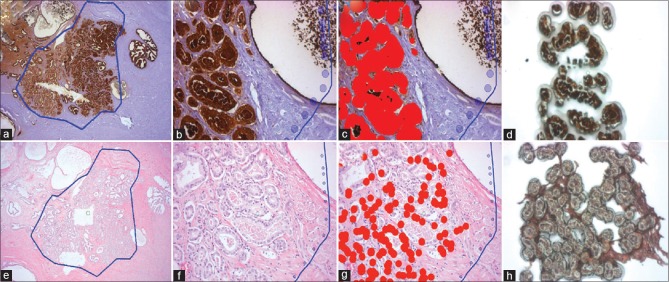
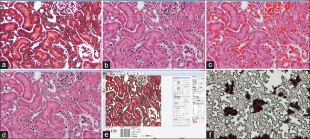
To improve the efficiency of the cellular identification process, we describe a microdissection workflow that leverages recently developed and open source image analysis algorithms referred to as computer-aided laser dissection (CALD). CALD permits a computer algorithm to identify the cells of interest and drive the dissection process.

RESULTS

We describe several "use cases" that demonstrate the integration of image analytic tools probabilistic pairwise Markov model, ImageJ, spatially invariant vector quantization (SIVQ), and eSeg onto the ThermoFisher Scientific ArcturusXT and Leica LMD7000 microdissection platforms.

CONCLUSIONS

The CALD methodology demonstrates the integration of image analysis tools with the microdissection workflow and shows the potential impact to clinical and life science applications.

摘要

引言

基于新一代测序和蛋白质组学的新型分子诊断检测方法的开发与应用,需要改进从组织切片中获取靶细胞的方法。激光显微切割能够基于视觉选择,成功地从组织标本中分离出不同的细胞,适用于许多研究和临床应用。然而,当分子分析需要大量细胞或需要评估相当数量的标本时,这可能是一项艰巨的任务。

材料与方法

为提高细胞识别过程的效率,我们描述了一种显微切割工作流程,该流程利用了最近开发的开源图像分析算法,即计算机辅助激光切割(CALD)。CALD允许计算机算法识别感兴趣的细胞并驱动切割过程。

结果

我们描述了几个“用例”,展示了图像分析工具概率成对马尔可夫模型、ImageJ、空间不变矢量量化(SIVQ)和eSeg在赛默飞世尔科技的ArcturusXT和徕卡LMD7000显微切割平台上的整合。

结论

CALD方法展示了图像分析工具与显微切割工作流程的整合,并显示了其对临床和生命科学应用的潜在影响。

https://cdn.ncbi.nlm.nih.gov/pmc/blobs/0e45/6298131/6c814f47a8ae/JPI-9-45-g006.jpg
https://cdn.ncbi.nlm.nih.gov/pmc/blobs/0e45/6298131/19682d9c88dc/JPI-9-45-g001.jpg
https://cdn.ncbi.nlm.nih.gov/pmc/blobs/0e45/6298131/9e26bcad5039/JPI-9-45-g002.jpg
https://cdn.ncbi.nlm.nih.gov/pmc/blobs/0e45/6298131/47f09eb4209e/JPI-9-45-g003.jpg
https://cdn.ncbi.nlm.nih.gov/pmc/blobs/0e45/6298131/dbf3552021f9/JPI-9-45-g004.jpg
https://cdn.ncbi.nlm.nih.gov/pmc/blobs/0e45/6298131/0c119af412b5/JPI-9-45-g005.jpg
https://cdn.ncbi.nlm.nih.gov/pmc/blobs/0e45/6298131/6c814f47a8ae/JPI-9-45-g006.jpg

相似文献

1
Computer-Aided Laser Dissection: A Microdissection Workflow Leveraging Image Analysis Tools.计算机辅助激光解剖:一种利用图像分析工具的显微解剖工作流程。
J Pathol Inform. 2018 Dec 11;9:45. doi: 10.4103/jpi.jpi_60_18. eCollection 2018.
2
SIVQ-LCM protocol for the ArcturusXT instrument.适用于ArcturusXT仪器的SIVQ-LCM方案。
J Vis Exp. 2014 Jul 23(89):51662. doi: 10.3791/51662.
3
SIVQ-aided laser capture microdissection: A tool for high-throughput expression profiling.SIVQ辅助激光捕获显微切割:一种用于高通量表达谱分析的工具。
J Pathol Inform. 2011 Mar 31;2:19. doi: 10.4103/2153-3539.78500.
4
Integration of architectural and cytologic driven image algorithms for prostate adenocarcinoma identification.基于架构和细胞驱动的图像算法融合用于前列腺腺癌识别。
Anal Cell Pathol (Amst). 2012;35(4):251-65. doi: 10.3233/ACP-2012-0054.
5
Spatially Invariant Vector Quantization: A pattern matching algorithm for multiple classes of image subject matter including pathology.空间不变矢量量化:一种用于包括病理学在内的多类图像主题的模式匹配算法。
J Pathol Inform. 2011 Feb 26;2:13. doi: 10.4103/2153-3539.77175.
6
Automated vector selection of SIVQ and parallel computing integration MATLAB™: Innovations supporting large-scale and high-throughput image analysis studies.SIVQ的自动向量选择与并行计算集成MATLAB™:支持大规模和高通量图像分析研究的创新方法。
J Pathol Inform. 2011;2:37. doi: 10.4103/2153-3539.83752. Epub 2011 Aug 13.
7
Image microarrays derived from tissue microarrays (IMA-TMA): New resource for computer-aided diagnostic algorithm development.源自组织微阵列的图像微阵列(IMA-TMA):用于计算机辅助诊断算法开发的新资源。
J Pathol Inform. 2012;3:24. doi: 10.4103/2153-3539.98168. Epub 2012 Jul 12.
8
Semiautomated laser capture microdissection of lung adenocarcinoma cytology samples.肺腺癌细胞学样本的半自动激光捕获显微切割
Acta Cytol. 2012;56(6):622-31. doi: 10.1159/000342984. Epub 2012 Nov 24.
9
Expression microdissection adapted to commercial laser dissection instruments.表达微切割适用于商业激光切割仪器。
Nat Protoc. 2011 Apr;6(4):457-67. doi: 10.1038/nprot.2010.202. Epub 2011 Mar 18.
10
Laser Microdissection Workflow for Isolating Nucleic Acids from Fixed and Frozen Tissue Samples.用于从固定和冷冻组织样本中分离核酸的激光显微切割工作流程。
Methods Mol Biol. 2018;1723:33-93. doi: 10.1007/978-1-4939-7558-7_3.

引用本文的文献

1
Image-Enabled Cell Sorting Using the BD CellView Technology.基于 BD CellView 技术的图像增强型细胞分选。
Methods Mol Biol. 2024;2779:145-158. doi: 10.1007/978-1-0716-3738-8_8.
2
Artificial intelligence-augmented histopathologic review using image analysis to optimize DNA yield from formalin-fixed paraffin-embedded slides.人工智能辅助的组织病理复习,使用图像分析从福尔马林固定石蜡包埋切片中优化 DNA 产量。
Mod Pathol. 2022 Dec;35(12):1791-1803. doi: 10.1038/s41379-022-01161-0. Epub 2022 Oct 5.
3
Effect of Antigen Retrieval on Genomic DNA From Immunodissected Samples.

本文引用的文献

1
Digital pathology image analysis: opportunities and challenges.数字病理学图像分析:机遇与挑战。
Imaging Med. 2009;1(1):7-10. doi: 10.2217/IIM.09.9.
2
Diagnostic Assessment of Deep Learning Algorithms for Detection of Lymph Node Metastases in Women With Breast Cancer.用于检测乳腺癌女性患者淋巴结转移的深度学习算法的诊断评估
JAMA. 2017 Dec 12;318(22):2199-2210. doi: 10.1001/jama.2017.14585.
3
An Image Analysis Resource for Cancer Research: PIIP-Pathology Image Informatics Platform for Visualization, Analysis, and Management.
抗原修复对免疫组化染色样本基因组 DNA 的影响
J Histochem Cytochem. 2022 Sep;70(9):643-658. doi: 10.1369/00221554221124163. Epub 2022 Sep 21.
4
Laser Capture Proteomics: spatial tissue molecular profiling from the bench to personalized medicine.激光捕获蛋白质组学:从实验台到个体化医学的空间组织分子剖析。
Expert Rev Proteomics. 2021 Oct;18(10):845-861. doi: 10.1080/14789450.2021.1984886. Epub 2021 Dec 14.
5
Application of Laser Microdissection to Uncover Regional Transcriptomics in Human Kidney Tissue.应用激光显微切割技术揭示人类肾脏组织中的区域转录组学
J Vis Exp. 2020 Jun 9(160). doi: 10.3791/61371.
癌症研究的图像分析资源:用于可视化、分析和管理的PIIP-病理学图像信息学平台。
Cancer Res. 2017 Nov 1;77(21):e83-e86. doi: 10.1158/0008-5472.CAN-17-0323.
4
Accurate and reproducible invasive breast cancer detection in whole-slide images: A Deep Learning approach for quantifying tumor extent.全切片图像中精确且可重复的浸润性乳腺癌检测:一种用于量化肿瘤范围的深度学习方法。
Sci Rep. 2017 Apr 18;7:46450. doi: 10.1038/srep46450.
5
A Deep Convolutional Neural Network for segmenting and classifying epithelial and stromal regions in histopathological images.一种用于在组织病理学图像中分割和分类上皮和基质区域的深度卷积神经网络。
Neurocomputing (Amst). 2016 May 26;191:214-223. doi: 10.1016/j.neucom.2016.01.034. Epub 2016 Feb 17.
6
Evaluating stability of histomorphometric features across scanner and staining variations: prostate cancer diagnosis from whole slide images.评估组织形态计量学特征在扫描仪和染色变化中的稳定性:基于全切片图像的前列腺癌诊断
J Med Imaging (Bellingham). 2016 Oct;3(4):047502. doi: 10.1117/1.JMI.3.4.047502. Epub 2016 Oct 24.
7
Deep learning for digital pathology image analysis: A comprehensive tutorial with selected use cases.用于数字病理学图像分析的深度学习:包含选定用例的全面教程。
J Pathol Inform. 2016 Jul 26;7:29. doi: 10.4103/2153-3539.186902. eCollection 2016.
8
Image analysis and machine learning in digital pathology: Challenges and opportunities.数字病理学中的图像分析与机器学习:挑战与机遇
Med Image Anal. 2016 Oct;33:170-175. doi: 10.1016/j.media.2016.06.037. Epub 2016 Jul 4.
9
Emerging Themes in Image Informatics and Molecular Analysis for Digital Pathology.数字病理学的图像信息学与分子分析中的新兴主题
Annu Rev Biomed Eng. 2016 Jul 11;18:387-412. doi: 10.1146/annurev-bioeng-112415-114722.
10
High-Throughput Microdissection for Next-Generation Sequencing.用于下一代测序的高通量显微切割
PLoS One. 2016 Mar 21;11(3):e0151775. doi: 10.1371/journal.pone.0151775. eCollection 2016.