Hosking Anna-Marie, Coakley Brandon J, Chang Dorothy, Talebi-Liasi Faezeh, Lish Samantha, Lee Sung Won, Zong Amanda M, Moore Ian, Browning James, Jacques Steven L, Krueger James G, Kelly Kristen M, Linden Kenneth G, Gareau Daniel S
Department of Dermatology, University of California Irvine, Irvine, California.
Laboratory for Investigative Dermatology, The Rockefeller University, New York, New York.
Lasers Surg Med. 2019 Mar;51(3):214-222. doi: 10.1002/lsm.23055. Epub 2019 Jan 17.
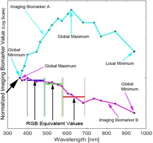
Early melanoma detection decreases morbidity and mortality. Early detection classically involves dermoscopy to identify suspicious lesions for which biopsy is indicated. Biopsy and histological examination then diagnose benign nevi, atypical nevi, or cancerous growths. With current methods, a considerable number of unnecessary biopsies are performed as only 11% of all biopsied, suspicious lesions are actually melanomas. Thus, there is a need for more advanced noninvasive diagnostics to guide the decision of whether or not to biopsy. Artificial intelligence can generate screening algorithms that transform a set of imaging biomarkers into a risk score that can be used to classify a lesion as a melanoma or a nevus by comparing the score to a classification threshold. Melanoma imaging biomarkers have been shown to be spectrally dependent in Red, Green, Blue (RGB) color channels, and hyperspectral imaging may further enhance diagnostic power. The purpose of this study was to use the same melanoma imaging biomarkers previously described, but over a wider range of wavelengths to determine if, in combination with machine learning algorithms, this could result in enhanced melanoma detection.
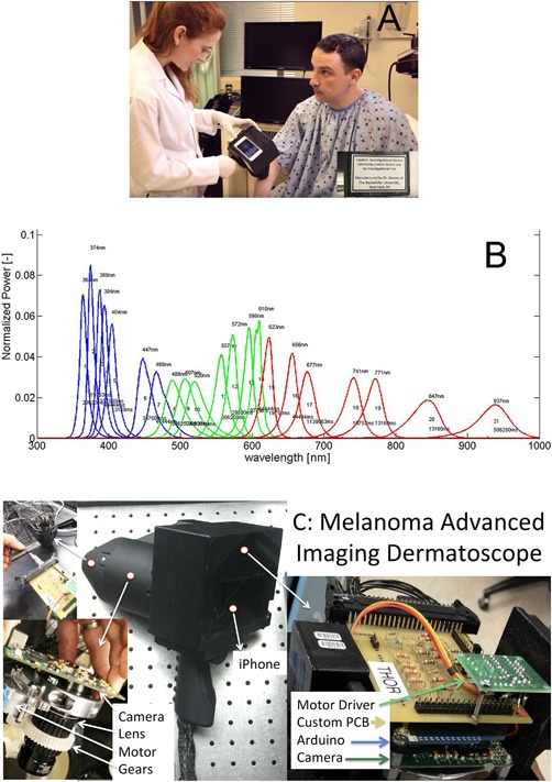
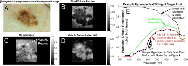
We used the melanoma advanced imaging dermatoscope (mAID) to image pigmented lesions assessed by dermatologists as requiring a biopsy. The mAID is a 21-wavelength imaging device in the 350-950 nm range. We then generated imaging biomarkers from these hyperspectral dermoscopy images, and, with the help of artificial intelligence algorithms, generated a melanoma Q-score for each lesion (0 = nevus, 1 = melanoma). The Q-score was then compared to the histopathologic diagnosis.
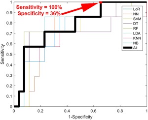
The overall sensitivity and specificity of hyperspectral dermoscopy in detecting melanoma when evaluated in a set of lesions selected by dermatologists as requiring biopsy was 100% and 36%, respectively.
With widespread application, and if validated in larger clinical trials, this non-invasive methodology could decrease unnecessary biopsies and potentially increase life-saving early detection events. Lasers Surg. Med. 51:214-222, 2019. © 2019 The Authors. Lasers in Surgery and Medicine Published by Wiley Periodicals, Inc.
早期黑色素瘤检测可降低发病率和死亡率。传统的早期检测方法包括皮肤镜检查,以识别需要进行活检的可疑病变。然后通过活检和组织学检查来诊断良性痣、非典型痣或癌性生长。采用目前的方法,大量活检是不必要的,因为在所有接受活检的可疑病变中,实际只有11%是黑色素瘤。因此,需要更先进的非侵入性诊断方法来指导是否进行活检的决策。人工智能可以生成筛查算法,将一组影像生物标志物转化为风险评分,通过将该评分与分类阈值进行比较,可用于将病变分类为黑色素瘤或痣。黑色素瘤影像生物标志物已被证明在红、绿、蓝(RGB)颜色通道中具有光谱依赖性,而高光谱成像可能会进一步提高诊断能力。本研究的目的是使用先前描述的相同黑色素瘤影像生物标志物,但在更宽的波长范围内,以确定结合机器学习算法是否能提高黑色素瘤的检测率。
我们使用黑色素瘤高级成像皮肤镜(mAID)对皮肤科医生评估为需要活检的色素沉着病变进行成像。mAID是一种波长范围在350 - 950nm的21波长成像设备。然后我们从这些高光谱皮肤镜图像中生成影像生物标志物,并借助人工智能算法为每个病变生成黑色素瘤Q评分(0 = 痣,1 = 黑色素瘤)。然后将Q评分与组织病理学诊断结果进行比较。
在皮肤科医生选择的一组需要活检的病变中评估时,高光谱皮肤镜检测黑色素瘤的总体敏感性和特异性分别为100%和36%。
随着广泛应用,并且如果在更大规模的临床试验中得到验证,这种非侵入性方法可能会减少不必要的活检,并有可能增加挽救生命的早期检测事件。《激光外科与医学》51:214 - 222,2019。© 2019作者。《激光外科与医学》由威利期刊公司出版。