• 文献检索
  • 文档翻译
  • 深度研究
  • 学术资讯
  • Suppr Zotero 插件Zotero 插件
  • 邀请有礼
  • 套餐&价格
  • 历史记录
应用&插件
Suppr Zotero 插件Zotero 插件浏览器插件Mac 客户端Windows 客户端微信小程序
定价
高级版会员购买积分包购买API积分包
服务
文献检索文档翻译深度研究API 文档MCP 服务
关于我们
关于 Suppr公司介绍联系我们用户协议隐私条款
关注我们

Suppr 超能文献

核心技术专利:CN118964589B侵权必究
粤ICP备2023148730 号-1Suppr @ 2026

文献检索

告别复杂PubMed语法,用中文像聊天一样搜索,搜遍4000万医学文献。AI智能推荐,让科研检索更轻松。

立即免费搜索

文件翻译

保留排版,准确专业,支持PDF/Word/PPT等文件格式,支持 12+语言互译。

免费翻译文档

深度研究

AI帮你快速写综述,25分钟生成高质量综述,智能提取关键信息,辅助科研写作。

立即免费体验

在健康和疾病状态下对脑部磁共振成像进行强大的、无图谱的自动分割。

Robust, atlas-free, automatic segmentation of brain MRI in health and disease.

作者信息

Selvaganesan Kartiga, Whitehead Emily, DeAlwis Paba M, Schindler Matthew K, Inati Souheil, Saad Ziad S, Ohayon Joan E, Cortese Irene C M, Smith Bryan, Nath Avindra, Reich Daniel S, Inati Sara, Nair Govind

机构信息

National Institute of Neurological Disorders and Stroke (NINDS), Bethesda, MD, 20893, USA.

Inati Analytics, Potomac, MD 20854, USA.

出版信息

Heliyon. 2019 Feb 18;5(2):e01226. doi: 10.1016/j.heliyon.2019.e01226. eCollection 2019 Feb.

DOI:10.1016/j.heliyon.2019.e01226
PMID:30828660
原文链接:https://pmc.ncbi.nlm.nih.gov/articles/PMC6383003/
Abstract

BACKGROUND

Brain- and lesion-volumes derived from magnetic resonance images (MRI) serve as important imaging markers of disease progression in neurodegenerative diseases and aging. While manual segmentation of these volumes is both tedious and impractical in large cohorts of subjects, automated segmentation methods often fail in accurate segmentation of brains with severe atrophy or high lesion loads. The purpose of this study was to develop an atlas-free brain Classification using DErivative-based Features (C-DEF), which utilizes all scans that may be acquired during the course of a routine MRI study at any center.

METHODS

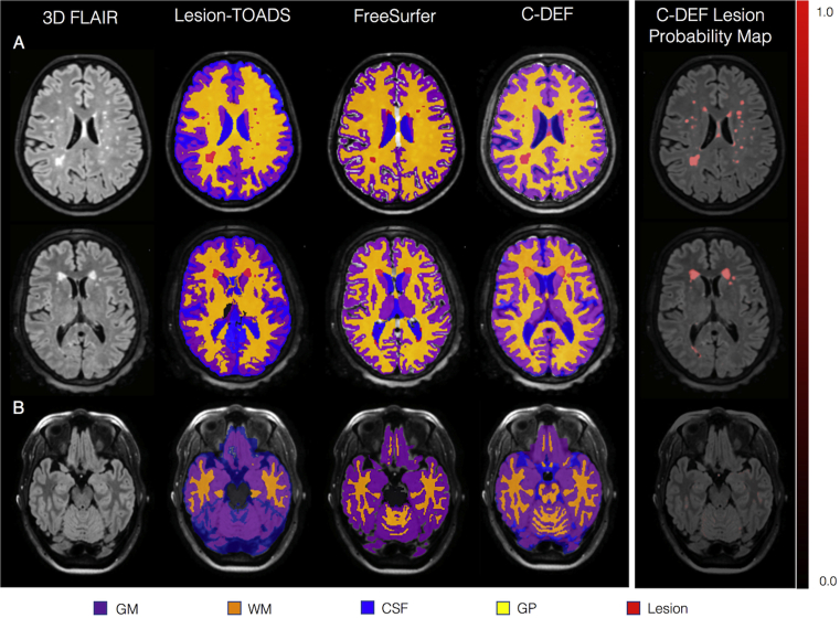
Proton-density, T-weighted, T-weighted, brain-free water, 3D FLAIR, 3D T-weighted, and 3D T*-weighted images, collected routinely on patients with neuroinflammatory diseases at the NIH, were used to optimize the C-DEF algorithm on healthy volunteers and HIV + subjects (cohort 1). First, manually marked lesions and eroded FreeSurfer brain segmentation masks (compiled into gray and white matter, globus pallidus, CSF labels) were used in training. Next, the optimized C-DEF was applied on a separate cohort of HIV + subjects (cohort two), and the results were compared with that of FreeSurfer and Lesion-TOADS. Finally, C-DEF segmentation was evaluated on subjects clinically diagnosed with various other neurological diseases (cohort three).

RESULTS

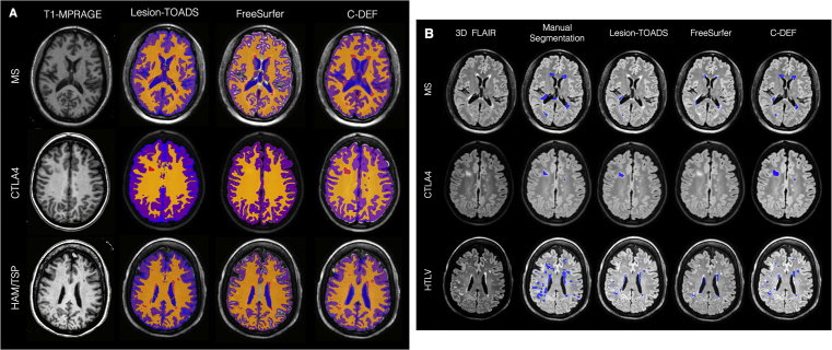
C-DEF algorithm was optimized using leave-one-out cross validation on five healthy subjects (age 36 ± 11 years), and five subjects infected with HIV (age 57 ± 2.6 years) in cohort one. The optimized C-DEF algorithm outperformed FreeSurfer and Lesion-TOADS segmentation in 49 other subjects infected with HIV (cohort two, age 54 ± 6 years) in qualitative and quantitative comparisons. Although trained only on HIV brains, sensitivity to detect lesions using C-DEF increased by 45% in HTLV-I-associated myelopathy/tropical spastic paraparesis (n = 5; age 58 ± 7 years), 33% in multiple sclerosis (n = 5; 42 ± 9 years old), and 4% in subjects with polymorphism of the cytotoxic T-lymphocyte-associated protein 4 gene (n = 5; age 24 ± 12 years) compared to Lesion-TOADS.

CONCLUSION

C-DEF outperformed other segmentation algorithms in the various neurological diseases explored herein, especially in lesion segmentation. While the results reported are from routine images acquired at the NIH, the algorithm can be easily trained and optimized for any set of contrasts and protocols for wider application. We are currently exploring various technical aspects of optimal implementation of CDEF in a clinical setting and evaluating a larger cohort of patients with other neurological diseases. Improving the accuracy of brain segmentation methodology will help better understand the relationship of imaging abnormalities to clinical and neuropsychological markers in disease.

摘要

背景

源自磁共振成像(MRI)的脑体积和病变体积是神经退行性疾病及衰老过程中疾病进展的重要影像标志物。虽然手动分割这些体积在大量受试者队列中既繁琐又不切实际,但自动分割方法在严重萎缩或病变负荷高的脑的准确分割中常常失败。本研究的目的是开发一种基于导数特征的无图谱脑分类法(C-DEF),该方法利用在任何中心的常规MRI研究过程中可能获取的所有扫描数据。

方法

美国国立卫生研究院(NIH)对患有神经炎性疾病的患者常规采集的质子密度、T加权、T加权、脑自由水、3D FLAIR、3D T加权和3D T*加权图像,用于在健康志愿者和HIV阳性受试者(队列1)上优化C-DEF算法。首先,在训练中使用手动标记的病变和侵蚀的FreeSurfer脑分割掩码(编译为灰质、白质、苍白球、脑脊液标签)。接下来,将优化后的C-DEF应用于另一组HIV阳性受试者(队列二),并将结果与FreeSurfer和Lesion-TOADS的结果进行比较。最后,在临床诊断为各种其他神经系统疾病的受试者(队列三)上评估C-DEF分割。

结果

在队列一中,使用留一法交叉验证对五名健康受试者(年龄36±11岁)和五名感染HIV的受试者(年龄57±2.6岁)优化了C-DEF算法。在定性和定量比较中,优化后的C-DEF算法在另外49名感染HIV的受试者(队列二,年龄54±6岁)中优于FreeSurfer和Lesion-TOADS分割。尽管仅在HIV脑上进行训练,但与Lesion-TOADS相比,使用C-DEF检测病变的敏感性在HTLV-I相关脊髓病/热带痉挛性截瘫(n = 5;年龄58±7岁)中提高了45%,在多发性硬化症(n = 5;42±9岁)中提高了33%,在细胞毒性T淋巴细胞相关蛋白4基因多态性受试者(n = 5;年龄24±12岁)中提高了4%。

结论

在本文探索的各种神经系统疾病中,C-DEF优于其他分割算法,尤其是在病变分割方面。虽然报告的结果来自于NIH采集的常规图像,但该算法可以很容易地针对任何一组对比和方案进行训练和优化,以实现更广泛的应用。我们目前正在探索CDEF在临床环境中最佳实施的各种技术方面,并评估更大队列的患有其他神经系统疾病的患者。提高脑分割方法的准确性将有助于更好地理解疾病中影像异常与临床和神经心理标志物之间的关系。

https://cdn.ncbi.nlm.nih.gov/pmc/blobs/51fe/6383003/bae595c621cd/gr7.jpg
https://cdn.ncbi.nlm.nih.gov/pmc/blobs/51fe/6383003/6b23246b634b/gr1.jpg
https://cdn.ncbi.nlm.nih.gov/pmc/blobs/51fe/6383003/67d154be1315/gr2.jpg
https://cdn.ncbi.nlm.nih.gov/pmc/blobs/51fe/6383003/8325bb36224b/gr3.jpg
https://cdn.ncbi.nlm.nih.gov/pmc/blobs/51fe/6383003/74d0cb49fea7/gr4.jpg
https://cdn.ncbi.nlm.nih.gov/pmc/blobs/51fe/6383003/108ef34ff332/gr5.jpg
https://cdn.ncbi.nlm.nih.gov/pmc/blobs/51fe/6383003/753b8ec71b4e/gr6.jpg
https://cdn.ncbi.nlm.nih.gov/pmc/blobs/51fe/6383003/bae595c621cd/gr7.jpg

相似文献

1
Robust, atlas-free, automatic segmentation of brain MRI in health and disease.在健康和疾病状态下对脑部磁共振成像进行强大的、无图谱的自动分割。
Heliyon. 2019 Feb 18;5(2):e01226. doi: 10.1016/j.heliyon.2019.e01226. eCollection 2019 Feb.
2
Logistic Regression-Based Model Is More Efficient Than U-Net Model for Reliable Whole Brain Magnetic Resonance Imaging Segmentation.基于逻辑回归的模型比 U-Net 模型更高效,可用于可靠的全脑磁共振成像分割。
Top Magn Reson Imaging. 2022 Jun 1;31(3):31-39. doi: 10.1097/RMR.0000000000000296. Epub 2022 Jun 28.
3
Automatic segmentation of white matter hyperintensities: validation and comparison with state-of-the-art methods on both Multiple Sclerosis and elderly subjects.自动分割脑白质高信号:在多发性硬化症和老年患者中验证和比较最先进的方法。
Neuroimage Clin. 2022;33:102940. doi: 10.1016/j.nicl.2022.102940. Epub 2022 Jan 10.
4
FLAIR-only joint volumetric analysis of brain lesions and atrophy in clinically isolated syndrome (CIS) suggestive of multiple sclerosis.FLAIR 仅用于联合评估疑似多发性硬化症的临床孤立综合征 (CIS) 患者的脑损伤和萎缩的容积分析。
Neuroimage Clin. 2021;29:102542. doi: 10.1016/j.nicl.2020.102542. Epub 2020 Dec 25.
5
Multi-atlas segmentation of the whole hippocampus and subfields using multiple automatically generated templates.使用多个自动生成的模板对整个海马体及其子区进行多图谱分割。
Neuroimage. 2014 Nov 1;101:494-512. doi: 10.1016/j.neuroimage.2014.04.054. Epub 2014 Apr 29.
6
Comparing lesion segmentation methods in multiple sclerosis: Input from one manually delineated subject is sufficient for accurate lesion segmentation.比较多发性硬化症中的病灶分割方法:一个手动勾画的病例输入就足以实现准确的病灶分割。
Neuroimage Clin. 2019;24:102074. doi: 10.1016/j.nicl.2019.102074. Epub 2019 Nov 5.
7
Evaluation of multimodal segmentation based on 3D T1-, T2- and FLAIR-weighted images - the difficulty of choosing.基于 3D T1、T2 和 FLAIR 加权图像的多模态分割评估——选择的困难。
Neuroimage. 2018 Apr 15;170:210-221. doi: 10.1016/j.neuroimage.2017.02.016. Epub 2017 Feb 7.
8
Performance of five research-domain automated WM lesion segmentation methods in a multi-center MS study.五种研究领域自动化 WM 病变分割方法在多中心 MS 研究中的性能。
Neuroimage. 2017 Dec;163:106-114. doi: 10.1016/j.neuroimage.2017.09.011. Epub 2017 Sep 9.
9
Human brain atlas for automated region of interest selection in quantitative susceptibility mapping: application to determine iron content in deep gray matter structures.人脑图谱用于定量磁化率映射中感兴趣区的自动选择:在确定深部灰质结构铁含量中的应用。
Neuroimage. 2013 Nov 15;82:449-69. doi: 10.1016/j.neuroimage.2013.05.127. Epub 2013 Jun 12.
10
Scanner agnostic large-scale evaluation of MS lesion delineation tool for clinical MRI.临床MRI中MS病变勾画工具的扫描仪无关大规模评估
Front Neurosci. 2023 May 19;17:1177540. doi: 10.3389/fnins.2023.1177540. eCollection 2023.

引用本文的文献

1
High-Field-Blinded Assessment of Portable Ultra-Low-Field Brain MRI for Multiple Sclerosis.便携式超低场脑磁共振成像对多发性硬化症的高场盲法评估
J Neuroimaging. 2025 Jan-Feb;35(1):e70005. doi: 10.1111/jon.70005.
2
Radiological Changes in the Spinal Cord and Brain of Patients with HTLV-1-Associated Myelopathy/Tropical Spastic Paraparesis (HAM/TSP).人类嗜T淋巴细胞病毒1型相关脊髓病/热带痉挛性截瘫(HAM/TSP)患者脊髓和脑部的放射学变化
Pathogens. 2024 Oct 22;13(11):920. doi: 10.3390/pathogens13110920.
3
Cardiovascular Disease, Brain Glucose Metabolism, and Neurocognitive Decline in People With Human Immunodeficiency Virus.

本文引用的文献

1
Neuroimaging markers of global cognition in early Alzheimer's disease: A magnetic resonance imaging-electroencephalography study.早期阿尔茨海默病患者全脑认知的神经影像学标志物:一项磁共振成像-脑电图研究。
Brain Behav. 2019 Jan;9(1):e01197. doi: 10.1002/brb3.1197. Epub 2018 Dec 27.
2
Atrophic brain signatures of mild forms of neurocognitive impairment in virally suppressed HIV infection.在病毒抑制的 HIV 感染中,轻度神经认知障碍的萎缩性脑特征。
AIDS. 2019 Jan 27;33(1):55-66. doi: 10.1097/QAD.0000000000002042.
3
Effect of interferon beta-1a subcutaneously three times weekly on clinical and radiological measures and no evidence of disease activity status in patients with relapsing-remitting multiple sclerosis at year 1.
人类免疫缺陷病毒感染者的心血管疾病、脑葡萄糖代谢与神经认知功能衰退
Open Forum Infect Dis. 2024 Sep 20;11(10):ofae552. doi: 10.1093/ofid/ofae552. eCollection 2024 Oct.
4
White and Gray Matter Changes are Associated With Neurocognitive Decline in HIV Infection.白质和灰质变化与HIV感染中的神经认知衰退相关。
Ann Neurol. 2024 May;95(5):941-950. doi: 10.1002/ana.26896. Epub 2024 Feb 16.
5
Pseudo-Label Assisted nnU-Net enables automatic segmentation of 7T MRI from a single acquisition.伪标签辅助nnU-Net可实现单次采集的7T磁共振成像自动分割。
Front Neuroimaging. 2023 Dec 1;2:1252261. doi: 10.3389/fnimg.2023.1252261. eCollection 2023.
6
Cortical lesions uniquely predict motor disability accrual and form rarely in the absence of new white matter lesions in multiple sclerosis.在多发性硬化症中,皮质病变独特地预示着运动功能障碍的累积,并且在没有新的白质病变的情况下很少出现。
medRxiv. 2023 Sep 25:2023.09.22.23295974. doi: 10.1101/2023.09.22.23295974.
7
Manual lesion segmentations for traumatic brain injury characterization.用于创伤性脑损伤特征描述的手动病变分割
Front Neuroimaging. 2023 Mar 16;2:1068591. doi: 10.3389/fnimg.2023.1068591. eCollection 2023.
8
Subject-Specific Automatic Reconstruction of White Matter Tracts.基于目标的白质束自动重建
J Digit Imaging. 2023 Dec;36(6):2648-2661. doi: 10.1007/s10278-023-00883-0. Epub 2023 Aug 3.
9
Advances in human intracranial electroencephalography research, guidelines and good practices.人类颅内脑电图研究进展、指南和良好实践。
Neuroimage. 2022 Oct 15;260:119438. doi: 10.1016/j.neuroimage.2022.119438. Epub 2022 Jul 2.
10
A Macaque Brain Extraction Model Based on U-Net Combined with Residual Structure.一种基于U-Net与残差结构相结合的猕猴脑提取模型。
Brain Sci. 2022 Feb 12;12(2):260. doi: 10.3390/brainsci12020260.
皮下注射干扰素β-1a每周三次对复发缓解型多发性硬化症患者1年时临床和影像学指标及无疾病活动证据状态的影响。
BMC Neurol. 2018 Sep 14;18(1):143. doi: 10.1186/s12883-018-1145-x.
4
Trial of Fingolimod versus Interferon Beta-1a in Pediatric Multiple Sclerosis.芬戈莫德与干扰素β-1a 在儿科多发性硬化症中的对比试验。
N Engl J Med. 2018 Sep 13;379(11):1017-1027. doi: 10.1056/NEJMoa1800149.
5
Neurofilament levels, disease activity and brain volume during follow-up in multiple sclerosis.多发性硬化症随访期间的神经丝水平、疾病活动和脑容量。
J Neuroinflammation. 2018 Jul 18;15(1):209. doi: 10.1186/s12974-018-1249-7.
6
White matter hyperintensities in relation to cognition in HIV-infected men with sustained suppressed viral load on combination antiretroviral therapy.接受联合抗逆转录病毒治疗且病毒载量持续受到抑制的HIV感染男性中,白质高信号与认知的关系
AIDS. 2016 Sep 24;30(15):2329-39. doi: 10.1097/QAD.0000000000001133.
7
Subject-Specific Sparse Dictionary Learning for Atlas-Based Brain MRI Segmentation.用于基于图谱的脑磁共振成像分割的特定主题稀疏字典学习
IEEE J Biomed Health Inform. 2015 Sep;19(5):1598-609. doi: 10.1109/JBHI.2015.2439242.
8
Association of Cortical Lesion Burden on 7-T Magnetic Resonance Imaging With Cognition and Disability in Multiple Sclerosis.7-T磁共振成像上皮质病变负荷与多发性硬化症认知及残疾的相关性
JAMA Neurol. 2015 Sep;72(9):1004-12. doi: 10.1001/jamaneurol.2015.1241.
9
Imaging endpoints for clinical trials in Alzheimer's disease.阿尔茨海默病临床试验的影像学终点指标。
Alzheimers Res Ther. 2014 Dec 20;6(9):87. doi: 10.1186/s13195-014-0087-9. eCollection 2014.
10
Quantitative Comparison of SPM, FSL, and Brainsuite for Brain MR Image Segmentation.用于脑磁共振图像分割的SPM、FSL和Brainsuite的定量比较。
J Biomed Phys Eng. 2014 Mar 8;4(1):13-26. eCollection 2014 Mar.