Section for Biomedical Imaging, University Medical Center Hamburg-Eppendorf, 20251, Hamburg, Germany.
Institute for Biomedical Imaging, Hamburg University of Technology, 21073, Hamburg, Germany.
Nat Commun. 2019 Apr 26;10(1):1936. doi: 10.1038/s41467-019-09704-x.
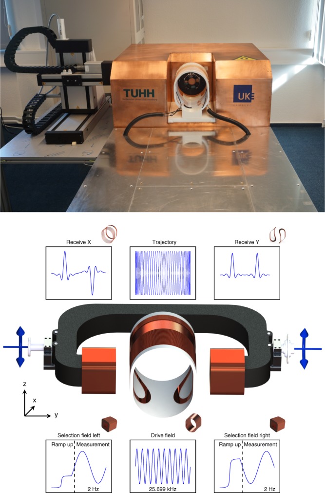
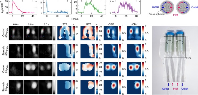
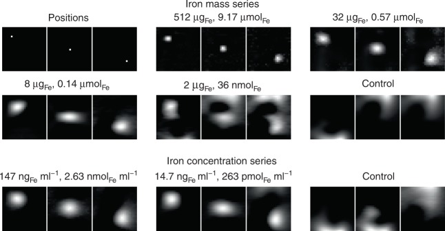
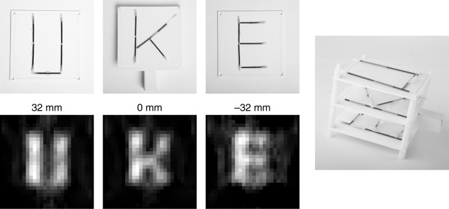
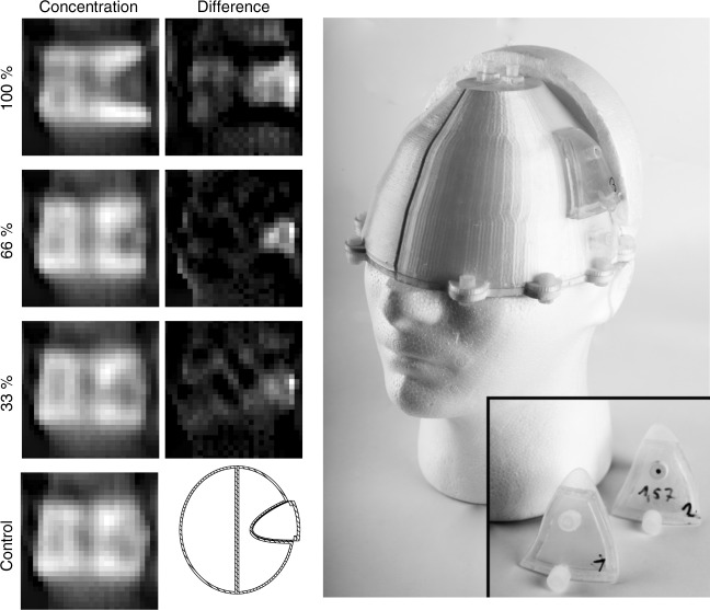
Determining the brain perfusion is an important task for diagnosis of vascular diseases such as occlusions and intracerebral haemorrhage. Even after successful diagnosis, there is a high risk of restenosis or rebleeding such that patients need intense attention in the days after treatment. Within this work, we present a diagnostic tomographic imager that allows access to brain perfusion quantitatively in short intervals. The device is based on the magnetic particle imaging technology and is designed for human scale. It is highly sensitive and allows the detection of an iron concentration of 263 pmol ml, which is one of the lowest iron concentrations imaged by MPI so far. The imager is self-shielded and can be used in unshielded environments such as intensive care units. In combination with the low technical requirements this opens up a variety of medical applications and would allow monitoring of stroke on intensive care units.
确定脑灌注是诊断血管疾病(如闭塞和脑出血)的重要任务。即使在成功诊断后,仍然存在很高的再狭窄或再出血风险,因此患者在治疗后几天需要密切关注。在这项工作中,我们提出了一种诊断层析成像仪,允许在短时间内定量获取脑灌注。该设备基于磁性粒子成像技术,设计用于人体尺度。它具有很高的灵敏度,允许检测到 263 pmol/ml 的铁浓度,这是迄今为止 MPI 成像的最低铁浓度之一。该成像仪具有自屏蔽功能,可以在重症监护病房等无屏蔽环境中使用。结合低技术要求,这为各种医疗应用开辟了道路,并允许在重症监护病房监测中风。