In Julie G, Foulke-Abel Jennifer, Clarke Elizabeth, Kovbasnjuk Olga
Department of Internal Medicine, University of New Mexico Health Science Center.
Department of Medicine, Division of Gastroenterology & Hepatology, Johns Hopkins University School of Medicine.
J Vis Exp. 2019 Apr 9(146). doi: 10.3791/59357.
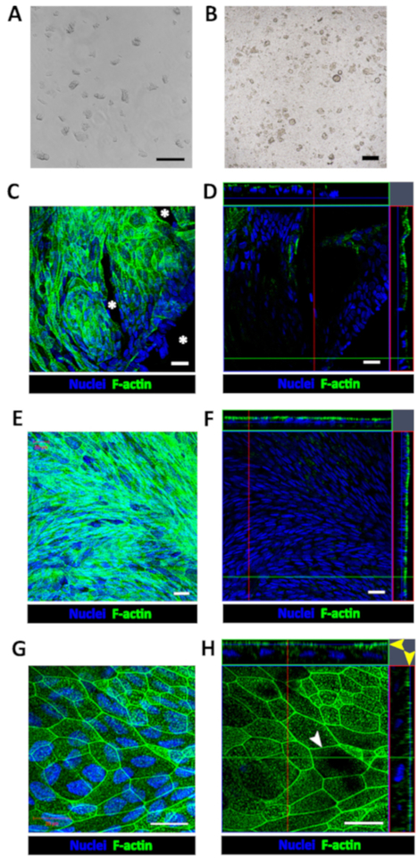
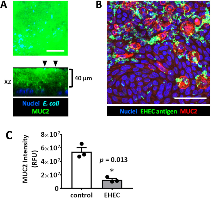
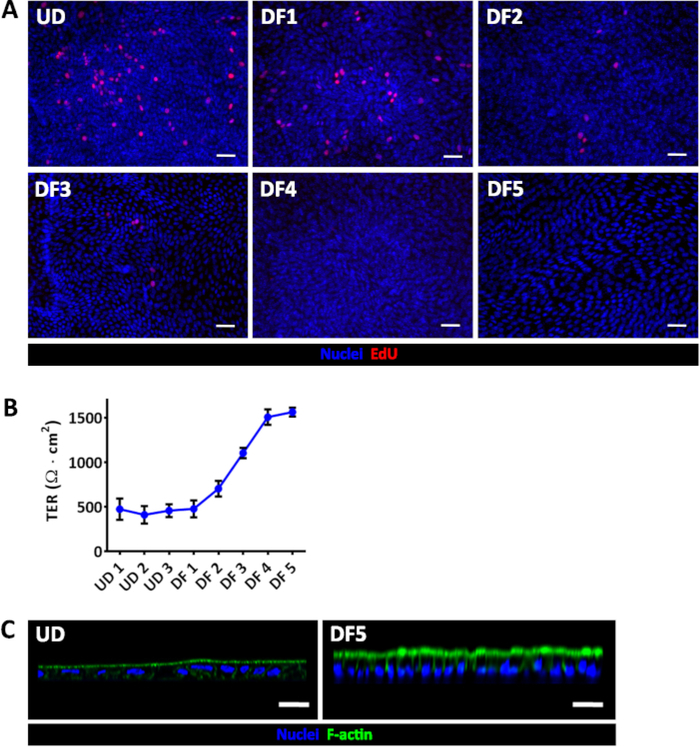
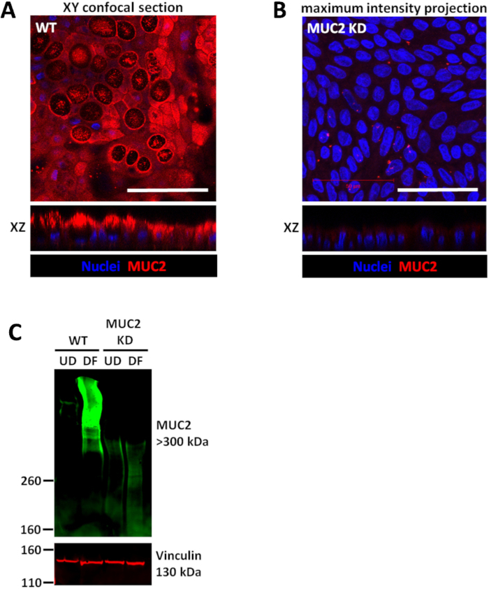
Human 3-dimensional (3D) enteroid or colonoid cultures derived from crypt base stem cells are currently the most advanced ex vivo model of the intestinal epithelium. Due to their closed structures and significant supporting extracellular matrix, 3D cultures are not ideal for host-pathogen studies. Enteroids or colonoids can be grown as epithelial monolayers on permeable tissue culture membranes to allow manipulation of both luminal and basolateral cell surfaces and accompanying fluids. This enhanced luminal surface accessibility facilitates modeling bacterial-host epithelial interactions such as the mucus-degrading ability of enterohemorrhagic E. coli (EHEC) on colonic epithelium. A method for 3D culture fragmentation, monolayer seeding, and transepithelial electrical resistance (TER) measurements to monitor the progress towards confluency and differentiation are described. Colonoid monolayer differentiation yields secreted mucus that can be studied by the immunofluorescence or immunoblotting techniques. More generally, enteroid or colonoid monolayers enable a physiologically-relevant platform to evaluate specific cell populations that may be targeted by pathogenic or commensal microbiota.
源自隐窝基部干细胞的人三维(3D)肠类器官或结肠类器官培养物是目前最先进的肠上皮体外模型。由于其封闭结构和大量支持性细胞外基质,3D培养物对于宿主-病原体研究并非理想选择。肠类器官或结肠类器官可以作为上皮单层在可渗透的组织培养膜上生长,以允许对管腔和基底外侧细胞表面以及伴随的液体进行操作。这种增强的管腔表面可及性有助于模拟细菌-宿主上皮相互作用,例如肠出血性大肠杆菌(EHEC)对结肠上皮的黏液降解能力。本文描述了一种用于3D培养物破碎、单层接种以及跨上皮电阻(TER)测量的方法,以监测融合和分化进程。结肠类器官单层分化产生可通过免疫荧光或免疫印迹技术研究的分泌黏液。更一般地说,肠类器官或结肠类器官单层提供了一个生理相关平台,用于评估可能被致病或共生微生物群靶向的特定细胞群体。