• 文献检索
  • 文档翻译
  • 深度研究
  • 学术资讯
  • Suppr Zotero 插件Zotero 插件
  • 邀请有礼
  • 套餐&价格
  • 历史记录
应用&插件
Suppr Zotero 插件Zotero 插件浏览器插件Mac 客户端Windows 客户端微信小程序
定价
高级版会员购买积分包购买API积分包
服务
文献检索文档翻译深度研究API 文档MCP 服务
关于我们
关于 Suppr公司介绍联系我们用户协议隐私条款
关注我们

Suppr 超能文献

核心技术专利:CN118964589B侵权必究
粤ICP备2023148730 号-1Suppr @ 2026

文献检索

告别复杂PubMed语法,用中文像聊天一样搜索,搜遍4000万医学文献。AI智能推荐,让科研检索更轻松。

立即免费搜索

文件翻译

保留排版,准确专业,支持PDF/Word/PPT等文件格式,支持 12+语言互译。

免费翻译文档

深度研究

AI帮你快速写综述,25分钟生成高质量综述,智能提取关键信息,辅助科研写作。

立即免费体验

利用切片排序平均和深度学习重建滤波器从同时多层 fMRI 数据中提取心电波。

Extraction of the cardiac waveform from simultaneous multislice fMRI data using slice sorted averaging and a deep learning reconstruction filter.

机构信息

Brain Imaging Center, McLean Hospital, 115 Mill Street, Belmont, MA, 02478, USA; Department of Psychiatry, Harvard University Medical School, Boston, MA, 02115, USA.

Brain Imaging Center, McLean Hospital, 115 Mill Street, Belmont, MA, 02478, USA; Department of Psychiatry, Harvard University Medical School, Boston, MA, 02115, USA.

出版信息

Neuroimage. 2019 Sep;198:303-316. doi: 10.1016/j.neuroimage.2019.05.049. Epub 2019 May 23.

DOI:10.1016/j.neuroimage.2019.05.049
PMID:31129302
原文链接:https://pmc.ncbi.nlm.nih.gov/articles/PMC6592732/
Abstract

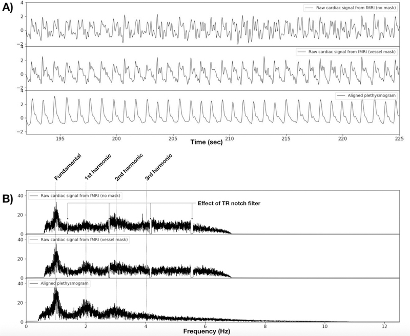
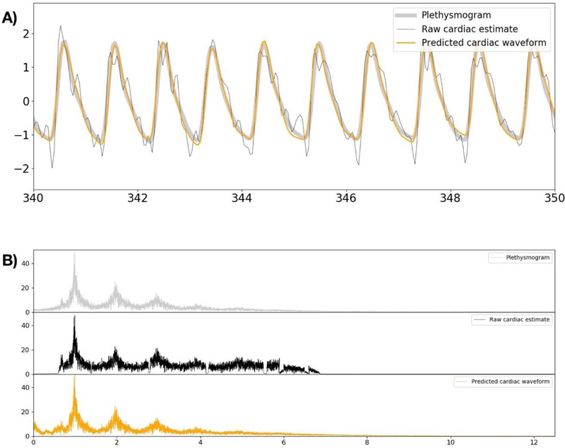
Cardiac signal contamination has long confounded the analysis of blood-oxygenation-level-dependent (BOLD) functional magnetic resonance imaging (fMRI). Cardiac pulsation results in significant BOLD signal changes, especially in and around blood vessels. Until the advent of simultaneous multislice echo-planar imaging (EPI) acquisition, the time resolution of whole brain EPI was insufficient to avoid cardiac aliasing (and acquisitions with repetition times (TRs) under 400-500 ms are still uncommon). As a result, direct detection and removal of the cardiac signal with spectral filters is generally not possible. Modelling methods have been developed to mitigate cardiac contamination, and recently developed techniques permit the visualization of cardiac signal propagation through the brain in undersampled data (e.g., TRs > 1s), which is useful in its own right for finding blood vessels. However, both of these techniques require data from which to estimate cardiac phase, which is generally not available for the data in many large databases of existing imaging data, and even now is not routinely recorded in many fMRI experiments. Here we present a method to estimate the cardiac waveform directly from a multislice fMRI dataset, without additional physiological measurements, such as plethysmograms. The pervasive spatial extent and temporal structure of the cardiac contamination signal across the brain offers an opportunity to exploit the nature of multislice imaging to extract this signal from the fMRI data itself. While any particular slice is recorded at the TR of the imaging experiment, slices are recorded much more quickly - typically from 10 to 20 Hz - sufficiently fast to fully sample the cardiac signal. Using the fairly permissive assumptions that the cardiac signal is a) pseudoperiodic b) somewhat coherent within any given slice, and c) is similarly shaped throughout the brain, we can extract a good estimate of the cardiac phase as a function of time from fMRI data alone. If we make further assumptions about the shape and consistency of cardiac waveforms, we can develop a deep learning filter to greatly improve our estimate of the cardiac waveform.

摘要

心脏信号污染长期以来一直困扰着血氧水平依赖(BOLD)功能磁共振成像(fMRI)的分析。心脏搏动会导致显著的 BOLD 信号变化,尤其是在血管内和周围。直到同时多层回波平面成像(EPI)采集技术问世之前,整个大脑 EPI 的时间分辨率不足以避免心脏伪影(重复时间(TR)低于 400-500ms 的采集仍然很少见)。因此,通常无法使用频谱滤波器直接检测和去除心脏信号。已经开发了建模方法来减轻心脏污染,最近开发的技术允许在欠采样数据中可视化心脏信号在大脑中的传播(例如,TR>1s),这本身对于发现血管就很有用。然而,这两种技术都需要估计心脏相位的数据,而许多现有的成像数据大型数据库中的数据通常没有这个相位的数据,即使在现在,许多 fMRI 实验也没有常规记录这个相位。在这里,我们提出了一种从多切片 fMRI 数据集直接估计心脏波形的方法,而无需额外的生理测量,如体积描记法。心脏污染信号在大脑中的普遍空间范围和时间结构为利用多切片成像的性质从 fMRI 数据本身中提取该信号提供了机会。虽然任何特定的切片都是在成像实验的 TR 记录的,但切片的记录速度要快得多-通常在 10 到 20Hz-足以对心脏信号进行完全采样。基于心脏信号是 a)准周期的、b)在任何给定切片内具有一定相干性、c)在整个大脑中具有相似形状的相当宽松的假设,我们可以仅从 fMRI 数据中提取出随时间变化的心脏相位的良好估计值。如果我们对心脏波形的形状和一致性做出进一步的假设,我们可以开发一种深度学习滤波器来大大提高我们对心脏波形的估计。

https://cdn.ncbi.nlm.nih.gov/pmc/blobs/f40d/6592732/1fbe88e7efb6/nihms-1530543-f0012.jpg
https://cdn.ncbi.nlm.nih.gov/pmc/blobs/f40d/6592732/68ec79058c1b/nihms-1530543-f0001.jpg
https://cdn.ncbi.nlm.nih.gov/pmc/blobs/f40d/6592732/4861f44b9dfb/nihms-1530543-f0002.jpg
https://cdn.ncbi.nlm.nih.gov/pmc/blobs/f40d/6592732/f839f95a85af/nihms-1530543-f0003.jpg
https://cdn.ncbi.nlm.nih.gov/pmc/blobs/f40d/6592732/9c635ec0123d/nihms-1530543-f0004.jpg
https://cdn.ncbi.nlm.nih.gov/pmc/blobs/f40d/6592732/c9b8063bf35f/nihms-1530543-f0005.jpg
https://cdn.ncbi.nlm.nih.gov/pmc/blobs/f40d/6592732/726b80dd8214/nihms-1530543-f0006.jpg
https://cdn.ncbi.nlm.nih.gov/pmc/blobs/f40d/6592732/b52ef37ee258/nihms-1530543-f0007.jpg
https://cdn.ncbi.nlm.nih.gov/pmc/blobs/f40d/6592732/89b0985e2b5d/nihms-1530543-f0008.jpg
https://cdn.ncbi.nlm.nih.gov/pmc/blobs/f40d/6592732/8becae584524/nihms-1530543-f0009.jpg
https://cdn.ncbi.nlm.nih.gov/pmc/blobs/f40d/6592732/1ab011fe25e3/nihms-1530543-f0010.jpg
https://cdn.ncbi.nlm.nih.gov/pmc/blobs/f40d/6592732/bf282bca3623/nihms-1530543-f0011.jpg
https://cdn.ncbi.nlm.nih.gov/pmc/blobs/f40d/6592732/1fbe88e7efb6/nihms-1530543-f0012.jpg

相似文献

1
Extraction of the cardiac waveform from simultaneous multislice fMRI data using slice sorted averaging and a deep learning reconstruction filter.利用切片排序平均和深度学习重建滤波器从同时多层 fMRI 数据中提取心电波。
Neuroimage. 2019 Sep;198:303-316. doi: 10.1016/j.neuroimage.2019.05.049. Epub 2019 May 23.
2
WHOCARES: WHOle-brain CArdiac signal REgression from highly accelerated simultaneous multi-Slice fMRI acquisitions.WHOCARES:从高度加速的同时多切片 fMRI 采集中回归全脑心脏信号。
J Neural Eng. 2022 Sep 6;19(5). doi: 10.1088/1741-2552/ac8bff.
3
Short repetition time multiband echo-planar imaging with simultaneous pulse recording allows dynamic imaging of the cardiac pulsation signal.具有同步脉冲记录功能的短重复时间多频段回波平面成像可实现心脏搏动信号的动态成像。
Magn Reson Med. 2014 Nov;72(5):1268-76. doi: 10.1002/mrm.25041. Epub 2013 Nov 22.
4
Prospective motion correction in functional MRI using simultaneous multislice imaging and multislice-to-volume image registration.前瞻性运动校正在功能磁共振成像中使用同时多切片成像和多切片到体积图像配准。
Neuroimage. 2019 Oct 15;200:159-173. doi: 10.1016/j.neuroimage.2019.06.042. Epub 2019 Jun 19.
5
Accelerated spin-echo functional MRI using multisection excitation by simultaneous spin-echo interleaving (MESSI) with complex-encoded generalized slice dithered enhanced resolution (cgSlider) simultaneous multislice echo-planar imaging.采用同时自旋回波交织多层面激发(MESSI)和复编码广义切片抖动增强分辨率(cgSlider)同时多层回波平面成像的加速自旋回波功能磁共振成像。
Magn Reson Med. 2020 Jul;84(1):206-220. doi: 10.1002/mrm.28108. Epub 2019 Dec 16.
6
Evaluation of 2D multiband EPI imaging for high-resolution, whole-brain, task-based fMRI studies at 3T: Sensitivity and slice leakage artifacts.3T场强下用于高分辨率、全脑、基于任务的功能磁共振成像研究的二维多频段回波平面成像评估:敏感性与层面间泄漏伪影
Neuroimage. 2016 Jan 1;124(Pt A):32-42. doi: 10.1016/j.neuroimage.2015.08.056. Epub 2015 Sep 1.
7
A circular echo planar sequence for fast volumetric fMRI.一种用于快速容积 fMRI 的圆形回波平面序列。
Magn Reson Med. 2019 Mar;81(3):1685-1698. doi: 10.1002/mrm.27522. Epub 2018 Oct 1.
8
Interleaved EPI based fMRI improved by multiplexed sensitivity encoding (MUSE) and simultaneous multi-band imaging.基于交错回波平面成像的功能磁共振成像通过多重灵敏度编码(MUSE)和同时多频段成像得到改进。
PLoS One. 2014 Dec 30;9(12):e116378. doi: 10.1371/journal.pone.0116378. eCollection 2014.
9
Generalized INverse imaging (GIN): ultrafast fMRI with physiological noise correction.广义逆成像(GIN):具有生理噪声校正功能的超快功能磁共振成像
Magn Reson Med. 2013 Oct;70(4):962-71. doi: 10.1002/mrm.24528. Epub 2012 Oct 24.
10
Rapid whole-brain resting-state fMRI at 3 T: Efficiency-optimized three-dimensional EPI versus repetition time-matched simultaneous-multi-slice EPI.3T 下快速全脑静息态 fMRI:效率优化的三维 EPI 与重复时间匹配的同时多层 EPI 比较。
Neuroimage. 2017 Dec;163:81-92. doi: 10.1016/j.neuroimage.2017.08.031. Epub 2017 Sep 18.

引用本文的文献

1
Functional MRI signals exhibit stronger covariation with peripheral autonomic measures as vigilance decreases.随着警觉性降低,功能磁共振成像信号与外周自主神经测量指标之间的协变更强。
Imaging Neurosci (Camb). 2024 Sep 13;2. doi: 10.1162/imag_a_00287. eCollection 2024.
2
Physiological Confounds in BOLD-fMRI and Their Correction.血氧水平依赖性功能磁共振成像中的生理干扰及其校正
NMR Biomed. 2025 Jul;38(7):e70076. doi: 10.1002/nbm.70076.
3
Sex-specific effects of intensity and dose of physical activity on BOLD-fMRI cerebrovascular reactivity and cerebral pulsatility.

本文引用的文献

1
Hypersampling of pseudo-periodic signals by analytic phase projection.通过解析相位投影对伪周期信号进行过采样。
Comput Biol Med. 2018 Jul 1;98:159-167. doi: 10.1016/j.compbiomed.2018.05.008. Epub 2018 May 16.
2
The resting-state fMRI arterial signal predicts differential blood transit time through the brain.静息态 fMRI 动脉信号可预测大脑中血液通过时间的差异。
J Cereb Blood Flow Metab. 2019 Jun;39(6):1148-1160. doi: 10.1177/0271678X17753329. Epub 2018 Jan 15.
3
Modeling Task fMRI Data Via Deep Convolutional Autoencoder.基于深度卷积自动编码器的功能磁共振成像数据建模。
体力活动强度和剂量对BOLD-fMRI脑血管反应性和脑搏动性的性别特异性影响。
J Cereb Blood Flow Metab. 2025 Mar 13:271678X251325399. doi: 10.1177/0271678X251325399.
4
Robust data-driven segmentation of pulsatile cerebral vessels using functional magnetic resonance imaging.利用功能磁共振成像对搏动性脑血管进行稳健的数据驱动分割
Interface Focus. 2024 Dec 6;14(6):20240024. doi: 10.1098/rsfs.2024.0024.
5
Pulsatility analysis of the circle of Willis.Willis环的搏动性分析
Aging Brain. 2024 Mar 9;5:100111. doi: 10.1016/j.nbas.2024.100111. eCollection 2024.
6
Unsupervised physiological noise correction of functional magnetic resonance imaging data using phase and magnitude information (PREPAIR).利用相位和幅度信息对功能磁共振成像数据进行无监督生理噪声校正(PREPAIR)。
Hum Brain Mapp. 2023 Feb 15;44(3):1209-1226. doi: 10.1002/hbm.26152. Epub 2022 Nov 19.
7
WHOCARES: WHOle-brain CArdiac signal REgression from highly accelerated simultaneous multi-Slice fMRI acquisitions.WHOCARES:从高度加速的同时多切片 fMRI 采集中回归全脑心脏信号。
J Neural Eng. 2022 Sep 6;19(5). doi: 10.1088/1741-2552/ac8bff.
8
Ultra-slow fMRI fluctuations in the fourth ventricle as a marker of drowsiness.第四脑室超慢 fMRI 波动可作为困倦的标志物。
Neuroimage. 2022 Oct 1;259:119424. doi: 10.1016/j.neuroimage.2022.119424. Epub 2022 Jun 30.
9
Coupling between cerebrovascular oscillations and CSF flow fluctuations during wakefulness: An fMRI study.清醒状态下脑血流波动与 CSF 流动波动的耦合:一项 fMRI 研究。
J Cereb Blood Flow Metab. 2022 Jun;42(6):1091-1103. doi: 10.1177/0271678X221074639. Epub 2022 Jan 16.
10
Imaging the temporal dynamics of brain states with highly sampled fMRI.利用高采样功能磁共振成像技术对大脑状态的时间动态进行成像。
Curr Opin Behav Sci. 2021 Aug;40:87-95. doi: 10.1016/j.cobeha.2021.02.005. Epub 2021 Mar 17.
IEEE Trans Med Imaging. 2018 Jul;37(7):1551-1561. doi: 10.1109/TMI.2017.2715285. Epub 2017 Jun 15.
4
Deep Convolutional Neural Networks for Image Classification: A Comprehensive Review.用于图像分类的深度卷积神经网络:全面综述
Neural Comput. 2017 Sep;29(9):2352-2449. doi: 10.1162/NECO_a_00990. Epub 2017 Jun 9.
5
The PhysIO Toolbox for Modeling Physiological Noise in fMRI Data.用于对功能磁共振成像数据中的生理噪声进行建模的生理工具箱
J Neurosci Methods. 2017 Jan 30;276:56-72. doi: 10.1016/j.jneumeth.2016.10.019. Epub 2016 Nov 8.
6
Correcting for Blood Arrival Time in Global Mean Regression Enhances Functional Connectivity Analysis of Resting State fMRI-BOLD Signals.在全局均值回归中校正血液到达时间可增强静息态功能磁共振成像血氧水平依赖信号的功能连接分析。
Front Hum Neurosci. 2016 Jun 28;10:311. doi: 10.3389/fnhum.2016.00311. eCollection 2016.
7
The first step for neuroimaging data analysis: DICOM to NIfTI conversion.神经影像数据分析的第一步:从DICOM格式转换为NIfTI格式。
J Neurosci Methods. 2016 May 1;264:47-56. doi: 10.1016/j.jneumeth.2016.03.001. Epub 2016 Mar 2.
8
Comparison of peripheral near-infrared spectroscopy low-frequency oscillations to other denoising methods in resting state functional MRI with ultrahigh temporal resolution.在超高时间分辨率静息态功能磁共振成像中,外周近红外光谱低频振荡与其他去噪方法的比较。
Magn Reson Med. 2016 Dec;76(6):1697-1707. doi: 10.1002/mrm.26038. Epub 2016 Feb 7.
9
Image Super-Resolution Using Deep Convolutional Networks.基于深度卷积网络的图像超分辨率重建。
IEEE Trans Pattern Anal Mach Intell. 2016 Feb;38(2):295-307. doi: 10.1109/TPAMI.2015.2439281.
10
Long-term neural and physiological phenotyping of a single human.对单个人类进行长期神经和生理表型分析。
Nat Commun. 2015 Dec 9;6:8885. doi: 10.1038/ncomms9885.