Department of Neurology, Massachusetts General Hospital, Harvard Medical School, Boston, MA, USA; Computer Science and Artificial Intelligence Lab, MIT, USA; Department of Population Health Sciences, German Centre for Neurodegenerative Diseases (DZNE), Germany.
Computer Science and Artificial Intelligence Lab, MIT, USA; Athinoula A. Martinos Center for Biomedical Imaging, Department of Radiology, Massachusetts General Hospital, Charlestown, MA, USA.
Neuroimage Clin. 2019;23:101884. doi: 10.1016/j.nicl.2019.101884. Epub 2019 May 29.
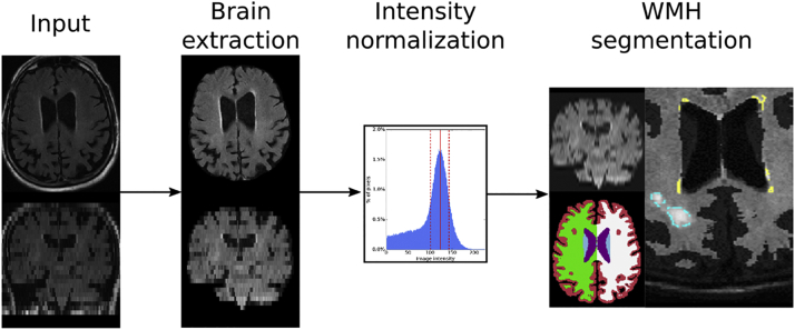
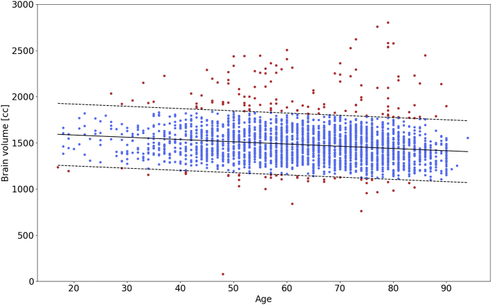
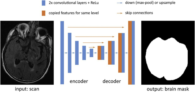
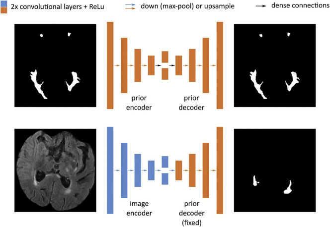
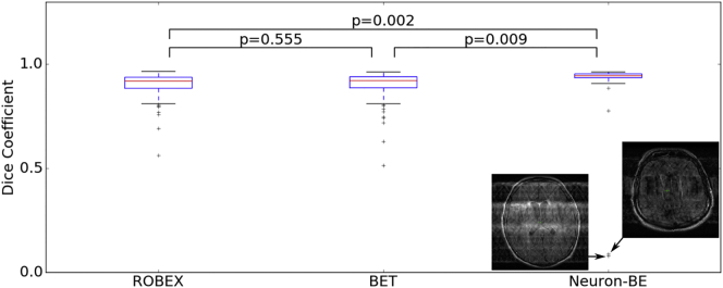
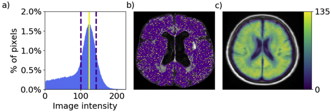
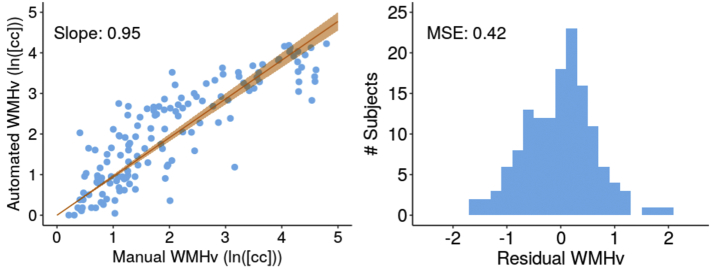
White matter hyperintensity (WMH) burden is a critically important cerebrovascular phenotype linked to prediction of diagnosis and prognosis of diseases, such as acute ischemic stroke (AIS). However, current approaches to its quantification on clinical MRI often rely on time intensive manual delineation of the disease on T2 fluid attenuated inverse recovery (FLAIR), which hinders high-throughput analyses such as genetic discovery. In this work, we present a fully automated pipeline for quantification of WMH in clinical large-scale studies of AIS. The pipeline incorporates automated brain extraction, intensity normalization and WMH segmentation using spatial priors. We first propose a brain extraction algorithm based on a fully convolutional deep learning architecture, specifically designed for clinical FLAIR images. We demonstrate that our method for brain extraction outperforms two commonly used and publicly available methods on clinical quality images in a set of 144 subject scans across 12 acquisition centers, based on dice coefficient (median 0.95; inter-quartile range 0.94-0.95; p < 0.01) and Pearson correlation of total brain volume (r = 0.90). Subsequently, we apply it to the large-scale clinical multi-site MRI-GENIE study (N = 2783) and identify a decrease in total brain volume of -2.4 cc/year. Additionally, we show that the resulting total brain volumes can successfully be used for quality control of image preprocessing. Finally, we obtain WMH volumes by building on an existing automatic WMH segmentation algorithm that delineates and distinguishes between different cerebrovascular pathologies. The learning method mimics expert knowledge of the spatial distribution of the WMH burden using a convolutional auto-encoder. This enables successful computation of WMH volumes of 2533 clinical AIS patients. We utilize these results to demonstrate the increase of WMH burden with age (0.950 cc/year) and show that single site estimates can be biased by the number of subjects recruited.
脑白质高信号(WMH)负担是一种与疾病的诊断和预后预测密切相关的重要脑血管表型,例如急性缺血性脑卒中(AIS)。然而,目前在临床 MRI 上对其进行量化的方法通常依赖于对 T2 液体衰减反转恢复(FLAIR)上疾病进行耗时的手动描绘,这阻碍了诸如遗传发现等高通量分析。在这项工作中,我们提出了一种用于在 AIS 的临床大规模研究中量化 WMH 的全自动流水线。该流水线结合了自动化的脑提取、强度归一化和基于空间先验的 WMH 分割。我们首先提出了一种基于全卷积深度学习架构的脑提取算法,该算法专门针对临床 FLAIR 图像设计。我们证明,在来自 12 个采集中心的 144 例患者扫描的一组图像上,与两种常用的、公开可用的方法相比,我们的脑提取方法在基于 Dice 系数(中位数 0.95;四分位距 0.94-0.95;p < 0.01)和总脑体积的 Pearson 相关系数(r = 0.90)方面表现更好。随后,我们将其应用于大型临床多中心 MRI-GENIE 研究(N = 2783),并发现总脑体积减少了 -2.4 cc/年。此外,我们表明,所得的总脑体积可以成功用于图像预处理的质量控制。最后,我们通过构建一个现有的自动 WMH 分割算法来获得 WMH 体积,该算法可以描绘和区分不同的脑血管病变。学习方法使用卷积自动编码器模仿 WMH 负担的空间分布的专家知识。这使得可以成功计算 2533 例临床 AIS 患者的 WMH 体积。我们利用这些结果来证明 WMH 负担随年龄增加(0.950 cc/年),并表明单个站点的估计可能会受到招募的受试者数量的影响。