• 文献检索
  • 文档翻译
  • 深度研究
  • 学术资讯
  • Suppr Zotero 插件Zotero 插件
  • 邀请有礼
  • 套餐&价格
  • 历史记录
应用&插件
Suppr Zotero 插件Zotero 插件浏览器插件Mac 客户端Windows 客户端微信小程序
定价
高级版会员购买积分包购买API积分包
服务
文献检索文档翻译深度研究API 文档MCP 服务
关于我们
关于 Suppr公司介绍联系我们用户协议隐私条款
关注我们

Suppr 超能文献

核心技术专利:CN118964589B侵权必究
粤ICP备2023148730 号-1Suppr @ 2026

文献检索

告别复杂PubMed语法,用中文像聊天一样搜索,搜遍4000万医学文献。AI智能推荐,让科研检索更轻松。

立即免费搜索

文件翻译

保留排版,准确专业,支持PDF/Word/PPT等文件格式,支持 12+语言互译。

免费翻译文档

深度研究

AI帮你快速写综述,25分钟生成高质量综述,智能提取关键信息,辅助科研写作。

立即免费体验

基于多尺度掩码区域卷积神经网络的PET成像肺部肿瘤检测

Multiscale Mask R-CNN-Based Lung Tumor Detection Using PET Imaging.

作者信息

Zhang Rui, Cheng Chao, Zhao Xuehua, Li Xuechen

机构信息

1 Department of Digital Media, Shenzhen Institute of Information Technology, Shenzhen, Guangdong, China.

2 Department of Nuclear Medicine, Changhai Hospital, Shanghai, People's Republic of China.

出版信息

Mol Imaging. 2019 Jan-Dec;18:1536012119863531. doi: 10.1177/1536012119863531.

DOI:10.1177/1536012119863531
PMID:31364467
原文链接:https://pmc.ncbi.nlm.nih.gov/articles/PMC6669841/
Abstract

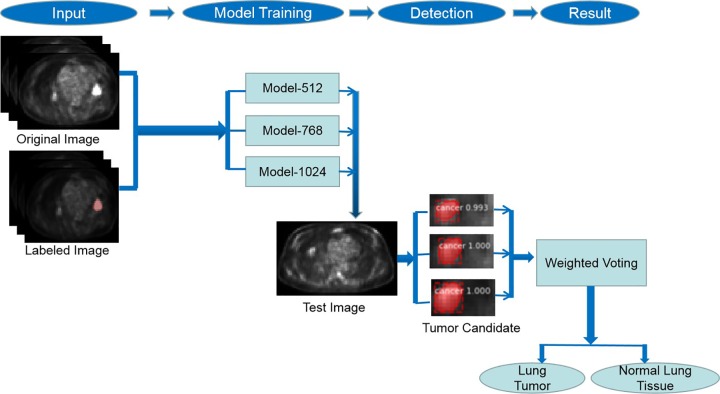
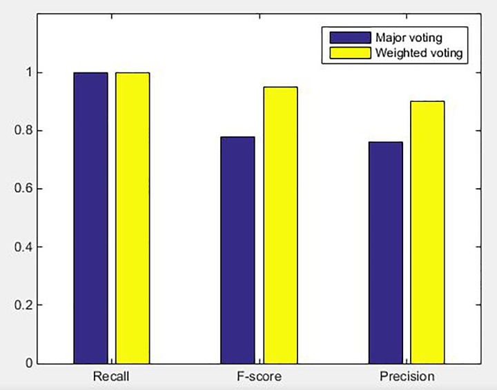
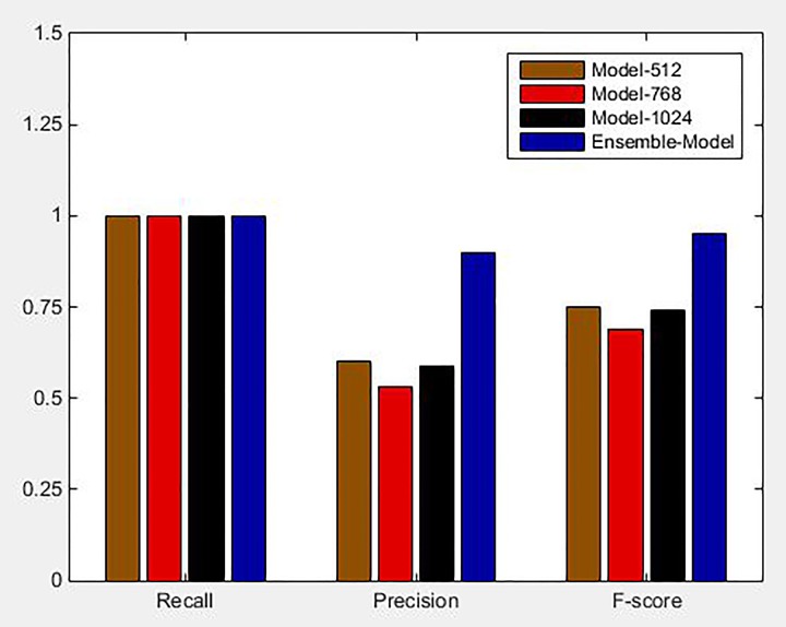
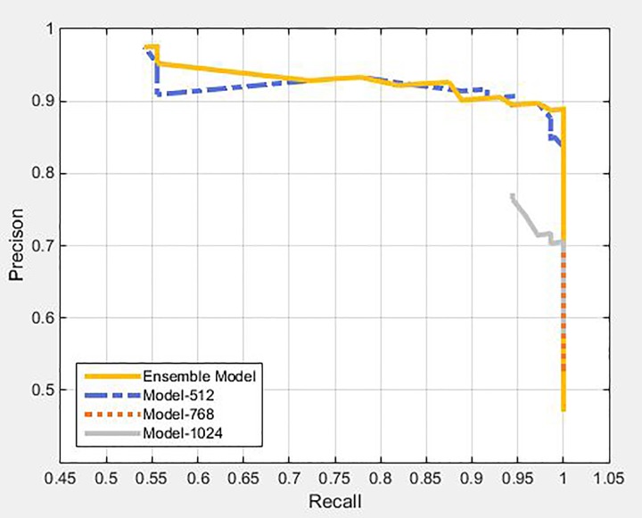
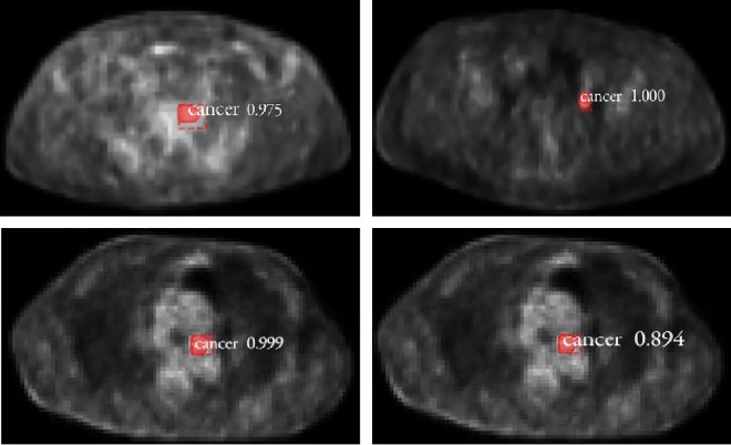
Positron emission tomography (PET) imaging serves as one of the most competent methods for the diagnosis of various malignancies, such as lung tumor. However, with an elevation in the utilization of PET scan, radiologists are overburdened considerably. Consequently, a new approach of "computer-aided diagnosis" is being contemplated to curtail the heavy workloads. In this article, we propose a multiscale Mask Region-Based Convolutional Neural Network (Mask R-CNN)-based method that uses PET imaging for the detection of lung tumor. First, we produced 3 models of Mask R-CNN for lung tumor candidate detection. These 3 models were generated by fine-tuning the Mask R-CNN using certain training data that consisted of images from 3 different scales. Each of the training data set included 594 slices with lung tumor. These 3 models of Mask R-CNN models were then integrated using weighted voting strategy to diminish the false-positive outcomes. A total of 134 PET slices were employed as test set in this experiment. The precision, recall, and score values of our proposed method were 0.90, 1, and 0.95, respectively. Experimental results exhibited strong conviction about the effectiveness of this method in detecting lung tumors, along with the capability of identifying a healthy chest pattern and reducing incorrect identification of tumors to a large extent.

摘要

正电子发射断层扫描(PET)成像作为诊断各种恶性肿瘤(如肺癌)最有效的方法之一。然而,随着PET扫描利用率的提高,放射科医生的负担大大加重。因此,正在考虑一种新的“计算机辅助诊断”方法来减轻繁重的工作量。在本文中,我们提出了一种基于多尺度基于区域的卷积神经网络(Mask R-CNN)的方法,该方法使用PET成像来检测肺癌。首先,我们生成了3个用于肺癌候选检测的Mask R-CNN模型。这3个模型是通过使用由3种不同尺度的图像组成的特定训练数据对Mask R-CNN进行微调而生成的。每个训练数据集包括594个有肺癌的切片。然后,这3个Mask R-CNN模型使用加权投票策略进行整合,以减少假阳性结果。在该实验中,总共使用了134个PET切片作为测试集。我们提出的方法的精确率、召回率和F1分数值分别为0.90、1和0.95。实验结果有力地证明了该方法在检测肺癌方面的有效性,以及识别健康胸部模式并在很大程度上减少肿瘤误判的能力。

https://cdn.ncbi.nlm.nih.gov/pmc/blobs/3a45/6669841/551673993d13/10.1177_1536012119863531-fig8.jpg
https://cdn.ncbi.nlm.nih.gov/pmc/blobs/3a45/6669841/ed893ba771f2/10.1177_1536012119863531-fig1.jpg
https://cdn.ncbi.nlm.nih.gov/pmc/blobs/3a45/6669841/b47b32feff08/10.1177_1536012119863531-fig2.jpg
https://cdn.ncbi.nlm.nih.gov/pmc/blobs/3a45/6669841/c25257e52572/10.1177_1536012119863531-fig3.jpg
https://cdn.ncbi.nlm.nih.gov/pmc/blobs/3a45/6669841/c380dee5c2fb/10.1177_1536012119863531-fig4.jpg
https://cdn.ncbi.nlm.nih.gov/pmc/blobs/3a45/6669841/9ad2d2f721f9/10.1177_1536012119863531-fig5.jpg
https://cdn.ncbi.nlm.nih.gov/pmc/blobs/3a45/6669841/0b905e423850/10.1177_1536012119863531-fig6.jpg
https://cdn.ncbi.nlm.nih.gov/pmc/blobs/3a45/6669841/1a22f5dc9131/10.1177_1536012119863531-fig7.jpg
https://cdn.ncbi.nlm.nih.gov/pmc/blobs/3a45/6669841/551673993d13/10.1177_1536012119863531-fig8.jpg

相似文献

1
Multiscale Mask R-CNN-Based Lung Tumor Detection Using PET Imaging.基于多尺度掩码区域卷积神经网络的PET成像肺部肿瘤检测
Mol Imaging. 2019 Jan-Dec;18:1536012119863531. doi: 10.1177/1536012119863531.
2
A CAD system for pulmonary nodule prediction based on deep three-dimensional convolutional neural networks and ensemble learning.基于深度三维卷积神经网络和集成学习的肺结节预测 CAD 系统。
PLoS One. 2019 Jul 12;14(7):e0219369. doi: 10.1371/journal.pone.0219369. eCollection 2019.
3
Automated detection of pulmonary nodules in PET/CT images: Ensemble false-positive reduction using a convolutional neural network technique.PET/CT图像中肺结节的自动检测:使用卷积神经网络技术减少总体假阳性
Med Phys. 2016 Jun;43(6):2821-2827. doi: 10.1118/1.4948498.
4
Generation of PET Attenuation Map for Whole-Body Time-of-Flight F-FDG PET/MRI Using a Deep Neural Network Trained with Simultaneously Reconstructed Activity and Attenuation Maps.基于同时重建的活性和衰减图训练的深度神经网络生成全身飞行时间 F-FDG PET/MRI 的 PET 衰减图。
J Nucl Med. 2019 Aug;60(8):1183-1189. doi: 10.2967/jnumed.118.219493. Epub 2019 Jan 25.
5
An effective approach for CT lung segmentation using mask region-based convolutional neural networks.基于掩模区域的卷积神经网络的 CT 肺分割有效方法。
Artif Intell Med. 2020 Mar;103:101792. doi: 10.1016/j.artmed.2020.101792. Epub 2020 Jan 8.
6
Automatic recognition of 3D GGO CT imaging signs through the fusion of hybrid resampling and layer-wise fine-tuning CNNs.通过混合重采样和逐层微调卷积神经网络融合实现 3D GGO CT 成像征象的自动识别。
Med Biol Eng Comput. 2018 Dec;56(12):2201-2212. doi: 10.1007/s11517-018-1850-z. Epub 2018 Jun 6.
7
[Computer aided diagnosis model for lung tumor based on ensemble convolutional neural network].基于集成卷积神经网络的肺肿瘤计算机辅助诊断模型
Sheng Wu Yi Xue Gong Cheng Xue Za Zhi. 2017 Aug 25;34(4):543-551. doi: 10.7507/1001-5515.201607003.
8
Single-view 2D CNNs with fully automatic non-nodule categorization for false positive reduction in pulmonary nodule detection.用于减少肺结节检测中假阳性的全自动无结节分类的单视图 2D CNN。
Comput Methods Programs Biomed. 2018 Oct;165:215-224. doi: 10.1016/j.cmpb.2018.08.012. Epub 2018 Aug 31.
9
Segmentation of lung parenchyma in CT images using CNN trained with the clustering algorithm generated dataset.基于聚类算法生成数据集训练的 CNN 对 CT 图像中的肺实质进行分割。
Biomed Eng Online. 2019 Jan 3;18(1):2. doi: 10.1186/s12938-018-0619-9.
10
Pear Recognition in an Orchard from 3D Stereo Camera Datasets to Develop a Fruit Picking Mechanism Using Mask R-CNN.从 3D 立体相机数据集识别梨树,以开发使用 Mask R-CNN 的果实采摘机制。
Sensors (Basel). 2022 May 31;22(11):4187. doi: 10.3390/s22114187.

引用本文的文献

1
Deep Learning Approaches for Automated Prediction of Treatment Response in Non-Small-Cell Lung Cancer Patients Based on CT and PET Imaging.基于CT和PET成像的非小细胞肺癌患者治疗反应自动预测的深度学习方法
Tomography. 2025 Jun 30;11(7):78. doi: 10.3390/tomography11070078.
2
Deep learning in nuclear medicine: from imaging to therapy.核医学中的深度学习:从成像到治疗。
Ann Nucl Med. 2025 May;39(5):424-440. doi: 10.1007/s12149-025-02031-w. Epub 2025 Mar 13.
3
PlaqueNet: deep learning enabled coronary artery plaque segmentation from coronary computed tomography angiography.

本文引用的文献

1
Mask R-CNN.Mask R-CNN。
IEEE Trans Pattern Anal Mach Intell. 2020 Feb;42(2):386-397. doi: 10.1109/TPAMI.2018.2844175. Epub 2018 Jun 5.
2
HEp-2 Specimen Image Segmentation and Classification Using Very Deep Fully Convolutional Network.使用深度全卷积网络进行 HEp-2 标本图像分割与分类。
IEEE Trans Med Imaging. 2017 Jul;36(7):1561-1572. doi: 10.1109/TMI.2017.2672702. Epub 2017 Feb 22.
3
Comparison of machine learning methods for classifying mediastinal lymph node metastasis of non-small cell lung cancer from F-FDG PET/CT images.
斑块网络:通过深度学习实现冠状动脉计算机断层扫描血管造影术对冠状动脉斑块的分割
Vis Comput Ind Biomed Art. 2024 Mar 22;7(1):6. doi: 10.1186/s42492-024-00157-8.
4
DraiNet: AI-driven decision support in pneumothorax and pleural effusion management.DraiNet:气胸和胸腔积液管理中的人工智能驱动决策支持。
Pediatr Surg Int. 2023 Dec 27;40(1):30. doi: 10.1007/s00383-023-05609-5.
5
An artificial intelligence method using FDG PET to predict treatment outcome in diffuse large B cell lymphoma patients.一种使用 FDG PET 的人工智能方法,用于预测弥漫性大 B 细胞淋巴瘤患者的治疗效果。
Sci Rep. 2023 Aug 12;13(1):13111. doi: 10.1038/s41598-023-40218-1.
6
Symmetrical awareness network for cross-site ultrasound thyroid nodule segmentation.用于跨站点超声甲状腺结节分割的对称感知网络。
Front Public Health. 2023 Mar 8;11:1055815. doi: 10.3389/fpubh.2023.1055815. eCollection 2023.
7
Automatic segmentation and measurement of pressure injuries using deep learning models and a LiDAR camera.利用深度学习模型和激光雷达相机进行压力性损伤的自动分割和测量。
Sci Rep. 2023 Jan 13;13(1):680. doi: 10.1038/s41598-022-26812-9.
8
Deep learning-assisted classification of calcaneofibular ligament injuries in the ankle joint.深度学习辅助诊断踝关节腓骨韧带损伤
Quant Imaging Med Surg. 2023 Jan 1;13(1):80-93. doi: 10.21037/qims-22-470. Epub 2022 Oct 13.
9
Analysis of Smart Lung Tumour Detector and Stage Classifier Using Deep Learning Techniques with Internet of Things.基于物联网的深度学习技术对智能肺部肿瘤探测器和分期分类器的分析。
Comput Intell Neurosci. 2022 Sep 13;2022:4608145. doi: 10.1155/2022/4608145. eCollection 2022.
10
Deep Learning-Based Internal Target Volume (ITV) Prediction Using Cone-Beam CT Images in Lung Stereotactic Body Radiotherapy.基于深度学习的肺立体定向体部放疗中锥形束 CT 图像的内部靶区(ITV)预测。
Technol Cancer Res Treat. 2022 Jan-Dec;21:15330338211073380. doi: 10.1177/15330338211073380.
基于F-FDG PET/CT图像的非小细胞肺癌纵隔淋巴结转移机器学习分类方法比较
EJNMMI Res. 2017 Dec;7(1):11. doi: 10.1186/s13550-017-0260-9. Epub 2017 Jan 28.
4
Single-Channel Sparse Non-Negative Blind Source Separation Method for Automatic 3-D Delineation of Lung Tumor in PET Images.单通道稀疏非负盲源分离方法在 PET 图像中自动勾画肺部肿瘤的三维边界
IEEE J Biomed Health Inform. 2017 Nov;21(6):1656-1666. doi: 10.1109/JBHI.2016.2624798. Epub 2016 Nov 3.
5
Faster R-CNN: Towards Real-Time Object Detection with Region Proposal Networks.更快的 R-CNN:基于区域建议网络的实时目标检测。
IEEE Trans Pattern Anal Mach Intell. 2017 Jun;39(6):1137-1149. doi: 10.1109/TPAMI.2016.2577031. Epub 2016 Jun 6.
6
Computer-aided diagnosis based on quantitative elastographic features with supersonic shear wave imaging.基于超声剪切波成像定量弹性特征的计算机辅助诊断
Ultrasound Med Biol. 2014 Feb;40(2):275-86. doi: 10.1016/j.ultrasmedbio.2013.09.032. Epub 2013 Nov 19.
7
Computer-aided diagnosis for phase-contrast X-ray computed tomography: quantitative characterization of human patellar cartilage with high-dimensional geometric features.相衬X射线计算机断层扫描的计算机辅助诊断:利用高维几何特征对人体髌软骨进行定量表征
J Digit Imaging. 2014 Feb;27(1):98-107. doi: 10.1007/s10278-013-9634-3.
8
Potential of computer-aided diagnosis of high spectral and spatial resolution (HiSS) MRI in the classification of breast lesions.高光谱和空间分辨率(HiSS)MRI 计算机辅助诊断在乳腺病变分类中的应用潜力。
J Magn Reson Imaging. 2014 Jan;39(1):59-67. doi: 10.1002/jmri.24145. Epub 2013 Sep 10.
9
Benefits and harms of CT screening for lung cancer: a systematic review.CT 筛查肺癌的获益与危害:系统评价。
JAMA. 2012 Jun 13;307(22):2418-29. doi: 10.1001/jama.2012.5521.
10
Automated radiation targeting in head-and-neck cancer using region-based texture analysis of PET and CT images.利用PET和CT图像的基于区域的纹理分析对头颈部癌进行自动放射靶向。
Int J Radiat Oncol Biol Phys. 2009 Oct 1;75(2):618-25. doi: 10.1016/j.ijrobp.2009.04.043. Epub 2009 Aug 14.