IEEE Trans Med Imaging. 2020 Mar;39(3):668-677. doi: 10.1109/TMI.2019.2933982. Epub 2019 Aug 8.
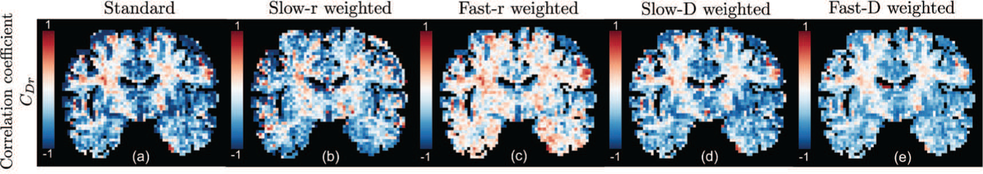
Joint relaxation-diffusion measurements can provide new insight about the tissue microstructural properties. Most recent methods have focused on inverting the Laplace transform to recover the joint distribution of relaxation-diffusion. However, as is well-known, this problem is notoriously ill-posed and numerically unstable. In this work, we address this issue by directly computing the joint moments of transverse relaxation rate and diffusivity, which can be robustly estimated. To zoom into different parts of the joint distribution, we further enhance our method by applying multiplicative filters to the joint probability density function of relaxation and diffusion and compute the corresponding moments. We propose an approach to use these moments to compute several novel scalar indices to characterize specific properties of the underlying tissue microstructure. Furthermore, for the first time, we propose an algorithm to estimate diffusion signals that are independent of echo time based on the moments of the marginal probability density function of diffusion. We demonstrate its utility in extracting tissue information not contaminated with multiple intra-voxel relaxation rates. We compare the performance of four types of filters that zoom into tissue components with different relaxation and diffusion properties and demonstrate it on an in-vivo human dataset. Experimental results show that these filters are able to characterize heterogeneous tissue microstructure. Moreover, the filtered diffusion signals are also able to distinguish fiber bundles with similar orientations but different relaxation rates. The proposed method thus allows to characterize the neural microstructure information in a robust and unique manner not possible using existing techniques.
关节弛豫-扩散测量可以提供关于组织微观结构特性的新见解。最近的方法主要集中在反演拉普拉斯变换以恢复弛豫-扩散的联合分布。然而,众所周知,这个问题是出了名的病态和数值不稳定的。在这项工作中,我们通过直接计算横向弛豫率和扩散的联合矩来解决这个问题,这些矩可以稳健地估计。为了放大联合分布的不同部分,我们通过对弛豫和扩散的联合概率密度函数应用乘法滤波器,并计算相应的矩,进一步增强了我们的方法。我们提出了一种方法,使用这些矩来计算几个新的标量指数,以表征基础组织微观结构的特定特性。此外,我们首次提出了一种基于扩散边际概率密度函数矩来估计不依赖回波时间的扩散信号的算法。我们在一个体内人类数据集上演示了它在提取不受多个体素内弛豫率污染的组织信息方面的效用。实验结果表明,这些滤波器能够描述具有不同弛豫和扩散特性的组织成分。此外,滤波后的扩散信号也能够区分具有相似取向但弛豫率不同的纤维束。因此,该方法允许以一种使用现有技术不可能的稳健且独特的方式来描述神经微观结构信息。