Department of Food Science and Human Nutrition, University of Illinois, Urbana, USA.
Department of Chemical and Biomolecular Engineering, University of Illinois, Urbana, USA.
Sci Rep. 2019 Aug 9;9(1):11560. doi: 10.1038/s41598-019-47903-0.
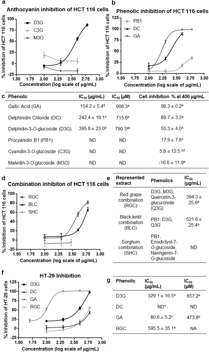
The objective was to assess anti-progression and stimulatory immune response effects among anthocyanins (ANC) and their metabolites on human colorectal cancer cells in vitro and in silico. Pure phenolics including delphinidin-3-O-glucoside (D3G) and its metabolites, delphinidin (DC) and gallic acid (GA), were tested alone or in combination, on HCT-116 and HT-29 human colorectal cancer cells (100-600 µg/mL). HCT-116 and HT-29 50% inhibition concentrations (µg/mL) were 396 ± 23 and 329 ± 17 for D3G; 242 ± 16 and >600 for DC; and 154 ± 5 and 81 ± 5 for GA, respectively. Using molecular docking, cyanidin-3-O-glucoside (C3G) showed the highest potential to inhibit immune checkpoints: programmed cell death protein-1 (PD-1) (-6.8 kcal/mol) and programmed death-ligand-1 (PD-L1) (-9.6 kcal/mol). C3G, D3G, DC, GA, and D3G-rich extracts decreased PD-L1 protein expression in HCT-116 cells. C3G decreased PD-L1 fluorescence intensity by 39%. ANC decreased PD-1 expression in peripheral blood mononuclear cells in monoculture by 41% and 55%, and co-culture with HCT-116 and HT-29 cells by 39% and 26% (C3G) and 50% and 51% (D3G), respectively. D3G and C3G, abundant in plant foods, showed potential for binding with and inhibiting immune checkpoints, PD-1 and PD-L1, which can activate immune response in the tumor microenvironment and induce cancer cell death.
目的是评估花色苷(ANC)及其代谢物在体外和体内对人结直肠癌细胞的抗进展和刺激免疫反应的作用。单独或联合测试了包括矢车菊素-3-O-葡萄糖苷(D3G)及其代谢物矢车菊素(DC)和没食子酸(GA)在内的纯酚类物质,对 HCT-116 和 HT-29 人结直肠癌细胞(100-600μg/ml)进行测试。HCT-116 和 HT-29 的 50%抑制浓度(μg/ml)分别为 396±23 和 329±17 对 D3G;242±16 和 >600 对 DC;154±5 和 81±5 对 GA。通过分子对接,矢车菊素-3-O-葡萄糖苷(C3G)显示出抑制免疫检查点:程序性细胞死亡蛋白-1(PD-1)(-6.8 kcal/mol)和程序性死亡配体-1(PD-L1)(-9.6 kcal/mol)的最大潜力。C3G、D3G、DC、GA 和富含 D3G 的提取物降低了 HCT-116 细胞中 PD-L1 蛋白的表达。C3G 使 PD-L1 荧光强度降低了 39%。ANC 在单核培养中降低了外周血单个核细胞中 PD-1 的表达 41%和 55%,与 HCT-116 和 HT-29 细胞共培养时降低了 39%和 26%(C3G)和 50%和 51%(D3G)。D3G 和 C3G,大量存在于植物性食物中,显示出与免疫检查点 PD-1 和 PD-L1 结合和抑制的潜力,这可以激活肿瘤微环境中的免疫反应并诱导癌细胞死亡。