• 文献检索
  • 文档翻译
  • 深度研究
  • 学术资讯
  • Suppr Zotero 插件Zotero 插件
  • 邀请有礼
  • 套餐&价格
  • 历史记录
应用&插件
Suppr Zotero 插件Zotero 插件浏览器插件Mac 客户端Windows 客户端微信小程序
定价
高级版会员购买积分包购买API积分包
服务
文献检索文档翻译深度研究API 文档MCP 服务
关于我们
关于 Suppr公司介绍联系我们用户协议隐私条款
关注我们

Suppr 超能文献

核心技术专利:CN118964589B侵权必究
粤ICP备2023148730 号-1Suppr @ 2026

文献检索

告别复杂PubMed语法,用中文像聊天一样搜索,搜遍4000万医学文献。AI智能推荐,让科研检索更轻松。

立即免费搜索

文件翻译

保留排版,准确专业,支持PDF/Word/PPT等文件格式,支持 12+语言互译。

免费翻译文档

深度研究

AI帮你快速写综述,25分钟生成高质量综述,智能提取关键信息,辅助科研写作。

立即免费体验

基于CT的头颈部鳞状细胞癌分级术前鉴别影像组学特征

CT-Based Radiomics Signature for the Preoperative Discrimination Between Head and Neck Squamous Cell Carcinoma Grades.

作者信息

Wu Wenli, Ye Junyong, Wang Qi, Luo Jin, Xu Shengsheng

机构信息

Department of Radiology, The First Affiliated Hospital, Chongqing Medical University, Chongqing, China.

Key Laboratory of Optoelectronic Technology and Systems of the Ministry of Education, Chongqing University, Chongqing, China.

出版信息

Front Oncol. 2019 Aug 30;9:821. doi: 10.3389/fonc.2019.00821. eCollection 2019.

DOI:10.3389/fonc.2019.00821
PMID:31544063
原文链接:https://pmc.ncbi.nlm.nih.gov/articles/PMC6729100/
Abstract

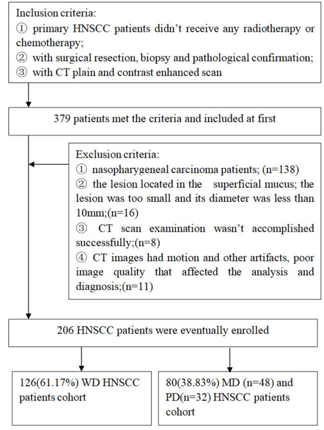
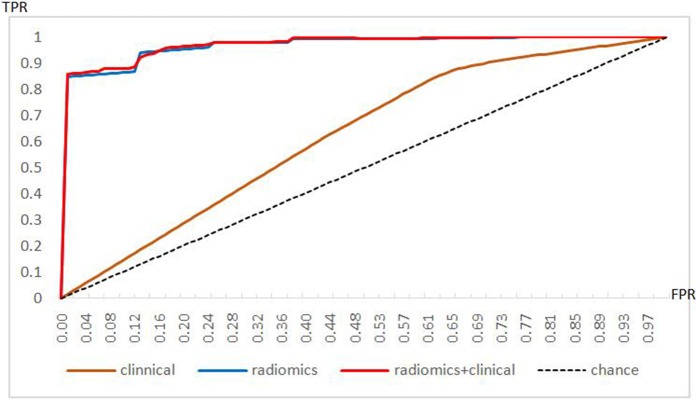
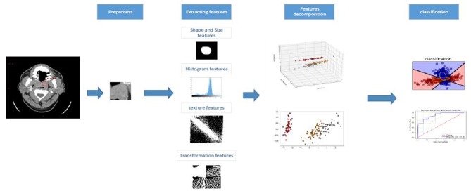
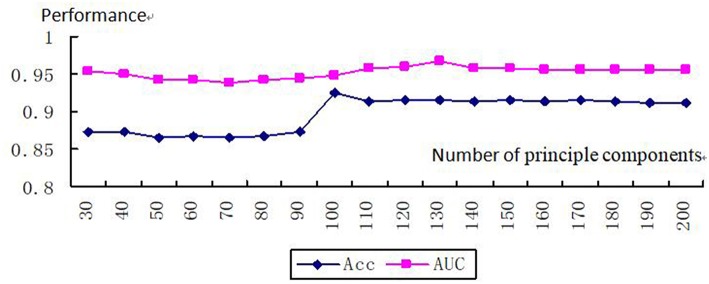
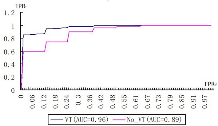
Radiomics has been widely used to non-invasively mine quantitative information from medical images and could potentially predict tumor phenotypes. Pathologic grade is considered a predictive prognostic factor for head and neck squamous cell carcinoma (HNSCC) patients. A preoperative histological assessment can be important in the clinical management of patients. We applied radiomics analysis to devise non-invasive biomarkers and accurately differentiate between well-differentiated (WD) and moderately differentiated (MD) and poorly differentiated (PD) HNSCC. This study involved 206 consecutive HNSCC patients (training cohort: = 137; testing cohort: = 69). In total, we extracted 670 radiomics features from contrast-enhanced computed tomography (CT) images. Radiomics signatures were constructed with a kernel principal component analysis (KPCA), random forest classifier and a variance-threshold (VT) selection. The associations between the radiomics signatures and HNSCC histological grades were investigated. A clinical model and combined model were also constructed. Areas under the receiver operating characteristic curves (AUCs) were applied to evaluate the performances of the three models. In total, 670 features were selected by the KPCA and random forest methods from the CT images. The radiomics signatures had a good performance in discriminating between the two cohorts of HNSCC grades, with an AUC of 0.96 and an accuracy of 0.92. The specificity, accuracy, sensitivity, positive predictive value (PPV), and negative predictive value (NPV) of the abovementioned method with a VT selection for determining HNSCC grades were 0.83, 0.92, 0.96, 0.94, and 0.91, respectively; without VT, the corresponding results were 0.70, 0.83, 0.88, 0.80, and 0.84. The differences in accuracy, sensitivity and NPV were significant between these approaches ( < 0.05). The AUCs with VT and without VT were 0.96 and 0.89, respectively ( < 0.05). Compared to the combined model and the radiomics signatures, The clinical model had a worse performance, and the differences were significant ( < 0.05). The combined model had the best performance, but the difference between the combined model and the radiomics signature weren't significant ( > 0.05). The CT-based radiomics signature could discriminate between WD and MD and PD HNSCC and might serve as a biomarker for preoperative grading.

摘要

放射组学已被广泛用于从医学图像中无创地挖掘定量信息,并有可能预测肿瘤表型。病理分级被认为是头颈部鳞状细胞癌(HNSCC)患者的一个预测性预后因素。术前组织学评估在患者的临床管理中可能很重要。我们应用放射组学分析来设计无创生物标志物,并准确区分高分化(WD)、中分化(MD)和低分化(PD)的HNSCC。本研究纳入了206例连续的HNSCC患者(训练队列: = 137;测试队列: = 69)。我们总共从增强计算机断层扫描(CT)图像中提取了670个放射组学特征。利用核主成分分析(KPCA)、随机森林分类器和方差阈值(VT)选择构建放射组学特征。研究了放射组学特征与HNSCC组织学分级之间的关联。还构建了一个临床模型和一个联合模型。应用受试者操作特征曲线(AUC)下的面积来评估这三个模型的性能。通过KPCA和随机森林方法从CT图像中总共选择了670个特征。放射组学特征在区分HNSCC分级的两个队列方面表现良好,AUC为0.96,准确率为0.92。上述采用VT选择来确定HNSCC分级的方法的特异性、准确率、敏感性、阳性预测值(PPV)和阴性预测值(NPV)分别为0.83、0.92、0.96、0.94和0.91;不采用VT时,相应结果分别为0.70、0.83、0.88、0.80和0.84。这些方法在准确率、敏感性和NPV方面的差异具有统计学意义(<0.05)。采用VT和不采用VT时的AUC分别为0.96和0.89(<0.05)。与联合模型和放射组学特征相比,临床模型的性能较差,差异具有统计学意义(<0.05)。联合模型表现最佳,但联合模型与放射组学特征之间的差异不具有统计学意义(>0.05)。基于CT的放射组学特征可以区分WD、MD和PD的HNSCC,并可能作为术前分级的生物标志物。

https://cdn.ncbi.nlm.nih.gov/pmc/blobs/f2f7/6729100/2939f8917dab/fonc-09-00821-g0006.jpg
https://cdn.ncbi.nlm.nih.gov/pmc/blobs/f2f7/6729100/a486a59370f2/fonc-09-00821-g0001.jpg
https://cdn.ncbi.nlm.nih.gov/pmc/blobs/f2f7/6729100/866b9b9b4a68/fonc-09-00821-g0002.jpg
https://cdn.ncbi.nlm.nih.gov/pmc/blobs/f2f7/6729100/cacd5e4a6033/fonc-09-00821-g0003.jpg
https://cdn.ncbi.nlm.nih.gov/pmc/blobs/f2f7/6729100/1728c19dceb8/fonc-09-00821-g0004.jpg
https://cdn.ncbi.nlm.nih.gov/pmc/blobs/f2f7/6729100/fa2dfa112e3a/fonc-09-00821-g0005.jpg
https://cdn.ncbi.nlm.nih.gov/pmc/blobs/f2f7/6729100/2939f8917dab/fonc-09-00821-g0006.jpg

相似文献

1
CT-Based Radiomics Signature for the Preoperative Discrimination Between Head and Neck Squamous Cell Carcinoma Grades.基于CT的头颈部鳞状细胞癌分级术前鉴别影像组学特征
Front Oncol. 2019 Aug 30;9:821. doi: 10.3389/fonc.2019.00821. eCollection 2019.
2
Magnetic resonance imaging based radiomics signature for the preoperative discrimination of stage I-II and III-IV head and neck squamous cell carcinoma.基于磁共振成像的放射组学特征用于术前鉴别 I-II 期和 III-IV 期头颈部鳞状细胞癌。
Eur J Radiol. 2018 Sep;106:1-6. doi: 10.1016/j.ejrad.2018.07.002. Epub 2018 Jul 4.
3
Dual-energy CT-based radiomics nomogram in predicting histological differentiation of head and neck squamous carcinoma: a multicenter study.基于双能 CT 的放射组学列线图预测头颈部鳞癌的组织学分化:一项多中心研究。
Neuroradiology. 2022 Feb;64(2):361-369. doi: 10.1007/s00234-021-02860-2. Epub 2021 Dec 3.
4
A CT-Based Deep Learning Radiomics Nomogram to Predict Histological Grades of Head and Neck Squamous Cell Carcinoma.基于 CT 的深度学习放射组学列线图预测头颈部鳞状细胞癌的组织学分级。
Acad Radiol. 2023 Aug;30(8):1591-1599. doi: 10.1016/j.acra.2022.11.007. Epub 2022 Nov 30.
5
A CT-based radiomics signature for preoperative discrimination between high and low expression of programmed death ligand 1 in head and neck squamous cell carcinoma.基于 CT 的放射组学特征用于术前鉴别头颈部鳞状细胞癌中程序性死亡配体 1 的高表达和低表达。
Eur J Radiol. 2022 Jan;146:110093. doi: 10.1016/j.ejrad.2021.110093. Epub 2021 Dec 4.
6
A computed tomography-based radiomics signature for predicting expression of programmed death ligand 1 in head and neck squamous cell carcinoma.基于计算机断层扫描的放射组学特征预测头颈部鳞状细胞癌程序性死亡配体 1 的表达。
Eur Radiol. 2022 Aug;32(8):5362-5370. doi: 10.1007/s00330-022-08651-4. Epub 2022 Mar 17.
7
Development and multicenter validation of a CT-based radiomics signature for discriminating histological grades of pancreatic ductal adenocarcinoma.基于CT的放射组学特征用于鉴别胰腺导管腺癌组织学分级的开发与多中心验证
Quant Imaging Med Surg. 2020 Mar;10(3):692-702. doi: 10.21037/qims.2020.02.21.
8
A multi-objective radiomics model for the prediction of locoregional recurrence in head and neck squamous cell cancer.一种用于预测头颈部鳞状细胞癌局部区域复发的多目标放射组学模型。
Med Phys. 2020 Oct;47(10):5392-5400. doi: 10.1002/mp.14388. Epub 2020 Aug 5.
9
Radiomics signature: a biomarker for the preoperative discrimination of lung invasive adenocarcinoma manifesting as a ground-glass nodule.影像组学特征:一种术前鉴别表现为磨玻璃结节的肺浸润性腺癌的生物标志物。
Eur Radiol. 2019 Feb;29(2):889-897. doi: 10.1007/s00330-018-5530-z. Epub 2018 Jul 2.
10
Application of contrast-enhanced CT radiomics in prediction of early recurrence of locally advanced oesophageal squamous cell carcinoma after trimodal therapy.增强 CT 影像组学在预测局部晚期食管鳞癌三模态治疗后早期复发中的应用。
Cancer Imaging. 2021 May 26;21(1):38. doi: 10.1186/s40644-021-00407-5.

引用本文的文献

1
MRI-based deep learning radiomics in predicting histological differentiation of oropharyngeal cancer: a multicenter cohort study.基于MRI的深度学习影像组学预测口咽癌组织学分化:一项多中心队列研究
Clin Transl Oncol. 2025 Sep 3. doi: 10.1007/s12094-025-04042-5.
2
Radiomics and radiogenomics: extracting more information from medical images for the diagnosis and prognostic prediction of ovarian cancer.放射组学和放射基因组学:从医学影像中提取更多信息用于卵巢癌的诊断和预后预测。
Mil Med Res. 2024 Dec 14;11(1):77. doi: 10.1186/s40779-024-00580-1.
3
Radiomics-based model for prediction of TGF-β1 expression in head and neck squamous cell carcinoma.

本文引用的文献

1
The utility of MRI histogram and texture analysis for the prediction of histological diagnosis in head and neck malignancies.MRI 直方图和纹理分析在头颈部恶性肿瘤组织学诊断预测中的应用。
Cancer Imaging. 2019 Feb 4;19(1):5. doi: 10.1186/s40644-019-0193-9.
2
Classification of the glioma grading using radiomics analysis.使用放射组学分析对胶质瘤进行分级分类。
PeerJ. 2018 Nov 22;6:e5982. doi: 10.7717/peerj.5982. eCollection 2018.
3
Quantitative parameters correlated well with differentiation of squamous cell carcinoma at head and neck: a study of dynamic contrast-enhanced MRI.
基于放射组学的头颈部鳞状细胞癌中转化生长因子-β1表达预测模型
Am J Nucl Med Mol Imaging. 2024 Aug 25;14(4):239-252. doi: 10.62347/JMKV7596. eCollection 2024.
4
Performance of radiomics in the differential diagnosis of parotid tumors: a systematic review.放射组学在腮腺肿瘤鉴别诊断中的应用:一项系统评价
Front Oncol. 2024 Jul 25;14:1383323. doi: 10.3389/fonc.2024.1383323. eCollection 2024.
5
Enhanced Diagnostic Precision: Assessing Tumor Differentiation in Head and Neck Squamous Cell Carcinoma Using Multi-Slice Spiral CT Texture Analysis.提高诊断精度:使用多层螺旋CT纹理分析评估头颈部鳞状细胞癌的肿瘤分化程度
J Clin Med. 2024 Jul 10;13(14):4038. doi: 10.3390/jcm13144038.
6
Artificial intelligence-driven radiomics study in cancer: the role of feature engineering and modeling.人工智能驱动的癌症放射组学研究:特征工程和建模的作用。
Mil Med Res. 2023 May 16;10(1):22. doi: 10.1186/s40779-023-00458-8.
7
Support Tools in the Differential Diagnosis of Salivary Gland Tumors through Inflammatory Biomarkers and Radiomics Metrics: A Preliminary Study.通过炎症生物标志物和放射组学指标进行唾液腺肿瘤鉴别诊断的支持工具:一项初步研究
Cancers (Basel). 2023 Mar 21;15(6):1876. doi: 10.3390/cancers15061876.
8
Development and validation of a CECT-based radiomics model for predicting IL1B expression and prognosis of head and neck squamous cell carcinoma.基于CT增强扫描的头颈部鳞状细胞癌IL1B表达及预后预测的影像组学模型的建立与验证
Front Oncol. 2023 Mar 10;13:1121485. doi: 10.3389/fonc.2023.1121485. eCollection 2023.
9
MRI Radiomics for Predicting Survival in Patients with Locally Advanced Hypopharyngeal Cancer Treated with Concurrent Chemoradiotherapy.磁共振成像放射组学预测局部晚期下咽癌同步放化疗患者的生存情况
Cancers (Basel). 2022 Dec 12;14(24):6119. doi: 10.3390/cancers14246119.
10
Utility of adding Radiomics to clinical features in predicting the outcomes of radiotherapy for head and neck cancer using machine learning.利用机器学习,放射组学在预测头颈部癌症放疗结果方面添加到临床特征中的效用。
PLoS One. 2022 Dec 15;17(12):e0277168. doi: 10.1371/journal.pone.0277168. eCollection 2022.
定量参数与头颈部鳞状细胞癌的分化密切相关:动态对比增强MRI研究
Acta Radiol. 2019 Aug;60(8):962-968. doi: 10.1177/0284185118809543. Epub 2018 Nov 20.
4
pN status predicts outcomes in surgically treated pT1-pT2 patients of various disease stages with squamous cell carcinoma of the head and neck: a 17-year retrospective single center cohort study.pN状态可预测手术治疗的不同疾病阶段的头颈部鳞状细胞癌pT1 - pT2患者的预后:一项为期17年的回顾性单中心队列研究。
Eur Arch Otorhinolaryngol. 2018 Nov;275(11):2787-2795. doi: 10.1007/s00405-018-5108-z. Epub 2018 Aug 29.
5
Magnetic resonance imaging based radiomics signature for the preoperative discrimination of stage I-II and III-IV head and neck squamous cell carcinoma.基于磁共振成像的放射组学特征用于术前鉴别 I-II 期和 III-IV 期头颈部鳞状细胞癌。
Eur J Radiol. 2018 Sep;106:1-6. doi: 10.1016/j.ejrad.2018.07.002. Epub 2018 Jul 4.
6
Diagnostic accuracy of MRI texture analysis for grading gliomas.MRI 纹理分析在胶质瘤分级中的诊断准确性。
J Neurooncol. 2018 Dec;140(3):583-589. doi: 10.1007/s11060-018-2984-4. Epub 2018 Aug 25.
7
Adverse pathologic features in T1/2 oral squamous cell carcinoma classified by the American Joint Committee on Cancer eighth edition and implications for treatment.美国癌症联合委员会第八版 T1/2 口腔鳞状细胞癌的不良病理特征分类及其对治疗的影响。
Head Neck. 2018 Oct;40(10):2123-2128. doi: 10.1002/hed.25168. Epub 2018 Aug 13.
8
Exploring Applications of Radiomics in Magnetic Resonance Imaging of Head and Neck Cancer: A Systematic Review.探索放射组学在头颈癌磁共振成像中的应用:一项系统综述。
Front Oncol. 2018 May 14;8:131. doi: 10.3389/fonc.2018.00131. eCollection 2018.
9
Histogram analysis parameters of apparent diffusion coefficient reflect tumor cellularity and proliferation activity in head and neck squamous cell carcinoma.表观扩散系数的直方图分析参数反映头颈部鳞状细胞癌的肿瘤细胞密度和增殖活性。
Oncotarget. 2018 May 4;9(34):23599-23607. doi: 10.18632/oncotarget.25284.
10
The value of histological grading of biopsy and resection specimens in early stage oral squamous cell carcinomas.早期口腔鳞状细胞癌活检和切除标本的组织学分级价值。
J Craniomaxillofac Surg. 2018 Jun;46(6):1001-1006. doi: 10.1016/j.jcms.2018.03.019. Epub 2018 Apr 5.