• 文献检索
  • 文档翻译
  • 深度研究
  • 学术资讯
  • Suppr Zotero 插件Zotero 插件
  • 邀请有礼
  • 套餐&价格
  • 历史记录
应用&插件
Suppr Zotero 插件Zotero 插件浏览器插件Mac 客户端Windows 客户端微信小程序
定价
高级版会员购买积分包购买API积分包
服务
文献检索文档翻译深度研究API 文档MCP 服务
关于我们
关于 Suppr公司介绍联系我们用户协议隐私条款
关注我们

Suppr 超能文献

核心技术专利:CN118964589B侵权必究
粤ICP备2023148730 号-1Suppr @ 2026

文献检索

告别复杂PubMed语法,用中文像聊天一样搜索,搜遍4000万医学文献。AI智能推荐,让科研检索更轻松。

立即免费搜索

文件翻译

保留排版,准确专业,支持PDF/Word/PPT等文件格式,支持 12+语言互译。

免费翻译文档

深度研究

AI帮你快速写综述,25分钟生成高质量综述,智能提取关键信息,辅助科研写作。

立即免费体验

超广角视网膜成像用于分析病理性近视类型与后巩膜葡萄肿之间的关联

Ultra-Widefield Retinal Imaging for Analyzing the Association Between Types of Pathological Myopia and Posterior Staphyloma.

作者信息

Mimura Ririko, Mori Kiwako, Torii Hidemasa, Nagai Norihiro, Suzuki Misa, Minami Sakiko, Ozawa Yoko, Kurihara Toshihide, Tsubota Kazuo

机构信息

Department of Ophthalmology, Ichikawa General Hospital, Tokyo Dental College, 5-11-13, Sugano, Ichikawa, Chiba 272-8513, Japan.

Department of Ophthalmology, Keio University School of Medicine, 35 Shinanomachi, Shinjuku-ku, Tokyo 160-8582, Japan.

出版信息

J Clin Med. 2019 Sep 20;8(10):1505. doi: 10.3390/jcm8101505.

DOI:10.3390/jcm8101505
PMID:31547036
原文链接:https://pmc.ncbi.nlm.nih.gov/articles/PMC6832756/
Abstract

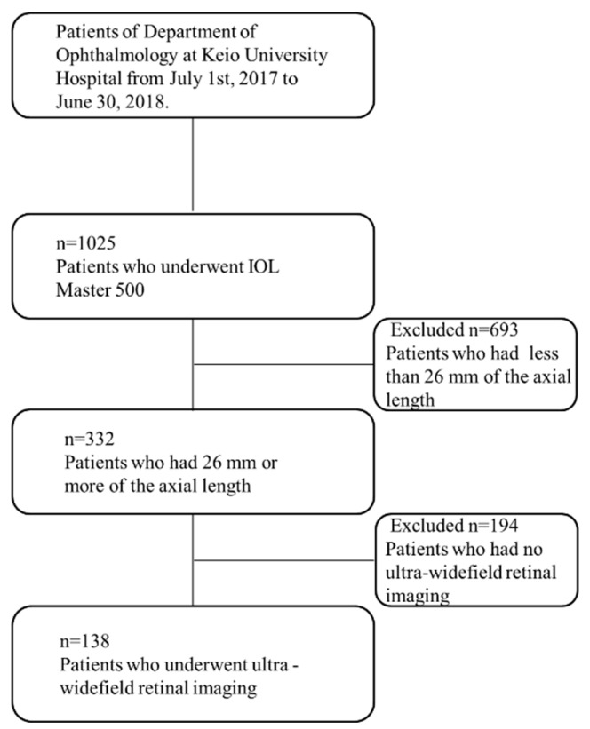
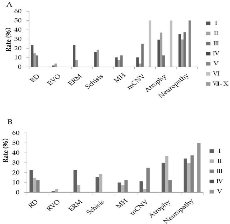
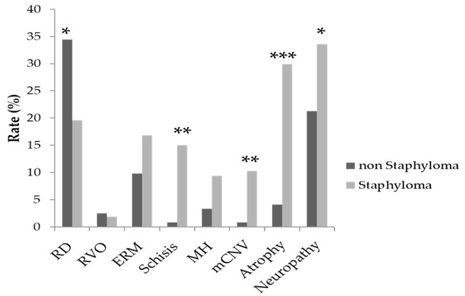
High myopia may develop to pathologic myopia, which brings severe visual impairment; however, the etiology is not fully understood. We, therefore, investigated the relationship between the presence of posterior staphyloma and posterior ocular disorders by assessing the patients with high myopia. A retrospective study was performed for the patients, who have more than 26 mm of the axial length and of whom fundus photography was taken with an ultra-widefield retinal imaging system. The objectives were 138 cases encompassing 229 eyes. In 138 cases, 91 were bilateral and 47 were unilateral. The averages ± SD of axial length of bilateral and unilateral were 28.8 ± 2.2 mm, 27.3 ± 1.2 mm, respectively, showing statistically significant difference. The number of eyes with and without posterior staphyloma were 107 (46.7%) and 122 (53.3%), respectively. Retinal detachment and retinal breaks are more observed in cases without posterior staphyloma ( = 0.017). Myopic choroidal neovascularization (mCNV) ( = 0.002), chorioretinal atrophy ( < 0.001), retinoschisis ( < 0.001), and optic neuropathy ( = 0.038) are more often seen in cases with posterior staphyloma. In conclusion, the prevalence rates of myopic choroidal neovascularization, retinal choroidal atrophy, and optic neuropathy were significantly higher with posterior staphyloma. The rate of periocular disorders such as retinal detachment was significantly higher without posterior staphyloma. These results indicate associations between types of pathological myopia and presence or absence of posterior staphyloma analyzed by ultra-widefield retinal imaging.

摘要

高度近视可能发展为病理性近视,从而导致严重的视力损害;然而,其病因尚未完全明确。因此,我们通过评估高度近视患者来研究后巩膜葡萄肿与后部眼部疾病之间的关系。对眼轴长度超过26mm且使用超广角视网膜成像系统进行眼底摄影的患者进行了一项回顾性研究。研究对象为138例患者,共229只眼。在138例患者中,91例为双眼,47例为单眼。双眼和单眼的眼轴长度平均值±标准差分别为28.8±2.2mm、27.3±1.2mm,差异具有统计学意义。有后巩膜葡萄肿和无后巩膜葡萄肿的眼数分别为107只(46.7%)和122只(53.3%)。视网膜脱离和视网膜裂孔在无后巩膜葡萄肿的病例中更为常见(P = 0.017)。近视性脉络膜新生血管(mCNV)(P = 0.002)、脉络膜视网膜萎缩(P < 0.001)、视网膜劈裂(P < 0.001)和视神经病变(P = 0.038)在有后巩膜葡萄肿的病例中更常出现。总之,后巩膜葡萄肿患者近视性脉络膜新生血管、视网膜脉络膜萎缩和视神经病变的患病率显著更高。无后巩膜葡萄肿时,视网膜脱离等眼周疾病的发生率显著更高。这些结果表明,通过超广角视网膜成像分析,病理性近视的类型与后巩膜葡萄肿的有无之间存在关联。

https://cdn.ncbi.nlm.nih.gov/pmc/blobs/17b6/6832756/5d2f952e7d8d/jcm-08-01505-g003.jpg
https://cdn.ncbi.nlm.nih.gov/pmc/blobs/17b6/6832756/3602b43f108d/jcm-08-01505-g001.jpg
https://cdn.ncbi.nlm.nih.gov/pmc/blobs/17b6/6832756/c227b241f8a5/jcm-08-01505-g002.jpg
https://cdn.ncbi.nlm.nih.gov/pmc/blobs/17b6/6832756/5d2f952e7d8d/jcm-08-01505-g003.jpg
https://cdn.ncbi.nlm.nih.gov/pmc/blobs/17b6/6832756/3602b43f108d/jcm-08-01505-g001.jpg
https://cdn.ncbi.nlm.nih.gov/pmc/blobs/17b6/6832756/c227b241f8a5/jcm-08-01505-g002.jpg
https://cdn.ncbi.nlm.nih.gov/pmc/blobs/17b6/6832756/5d2f952e7d8d/jcm-08-01505-g003.jpg

相似文献

1
Ultra-Widefield Retinal Imaging for Analyzing the Association Between Types of Pathological Myopia and Posterior Staphyloma.超广角视网膜成像用于分析病理性近视类型与后巩膜葡萄肿之间的关联
J Clin Med. 2019 Sep 20;8(10):1505. doi: 10.3390/jcm8101505.
2
Role of Ultra-widefield Imaging in the evaluation of Long-term change of highly myopic fundus.超广角成像在高度近视眼底长期变化评估中的作用。
Acta Ophthalmol. 2022 Jun;100(4):e977-e985. doi: 10.1111/aos.15009. Epub 2021 Aug 17.
3
CLINICAL AND MORPHOLOGIC FEATURES OF POSTERIOR STAPHYLOMA EDGES BY ULTRA-WIDEFIELD IMAGING IN PATHOLOGIC MYOPIA.病理性近视超广角成像后葡萄肿边缘的临床及形态学特征。
Retina. 2021 Nov 1;41(11):2278-2287. doi: 10.1097/IAE.0000000000003231.
4
Myopia-related fundus changes in Singapore adults with high myopia.新加坡高度近视成人的近视相关眼底改变。
Am J Ophthalmol. 2013 Jun;155(6):991-999.e1. doi: 10.1016/j.ajo.2013.01.016. Epub 2013 Mar 15.
5
Prevalence and characteristics of foveal retinal detachment without macular hole in high myopia.高度近视性无视网膜裂孔的黄斑区视网膜脱离的患病率及特征
Am J Ophthalmol. 2003 Mar;135(3):338-42. doi: 10.1016/s0002-9394(02)01937-2.
6
Clinical Characteristics of Posterior Staphylomas in Myopic Eyes With Axial Length Shorter Than 26.5 Millimeters.眼轴长度短于26.5毫米的近视眼中后巩膜葡萄肿的临床特征
Am J Ophthalmol. 2016 Feb;162:180-190.e1. doi: 10.1016/j.ajo.2015.11.016. Epub 2015 Nov 14.
7
Prevalence and Associations of Posterior Segment Manifestations in a Cohort of Egyptian Patients with Pathological Myopia.埃及病理性近视患者队列中后段表现的患病率及相关因素。
Curr Eye Res. 2019 Sep;44(9):955-962. doi: 10.1080/02713683.2019.1606252. Epub 2019 Apr 30.
8
Myopic maculopathy: Current status and proposal for a new classification and grading system (ATN).近视性黄斑病变:现状与新分类分级系统(ATN)的建议
Prog Retin Eye Res. 2019 Mar;69:80-115. doi: 10.1016/j.preteyeres.2018.10.005. Epub 2018 Nov 1.
9
Three-Dimensional Eye Shape, Myopic Maculopathy, and Visual Acuity: The Zhongshan Ophthalmic Center-Brien Holden Vision Institute High Myopia Cohort Study.三维眼形、近视性黄斑病变和视力:中山眼科中心-布莱恩·霍顿视觉研究所高度近视队列研究。
Ophthalmology. 2017 May;124(5):679-687. doi: 10.1016/j.ophtha.2017.01.009. Epub 2017 Feb 23.
10
Macular complications associated with posterior staphyloma.与后巩膜葡萄肿相关的黄斑并发症。
Am J Ophthalmol. 1997 Feb;123(2):181-7. doi: 10.1016/s0002-9394(14)71034-7.

引用本文的文献

1
Comprehensive analysis of posterior segment characteristics in high myopic Turkish patients.对高度近视土耳其患者眼后段特征的综合分析。
BMC Ophthalmol. 2025 Jun 3;25(1):335. doi: 10.1186/s12886-025-04167-9.
2
Thicker peripapillary choroid may be associated with behavioral factors in Tokyo's children.较厚的视乳头周围脉络膜可能与东京儿童的行为因素有关。
Sci Rep. 2025 May 22;15(1):17868. doi: 10.1038/s41598-025-02825-y.
3
Choroidal thinning in myopia is associated with axial elongation and severity of myopic maculopathy.近视患者的脉络膜变薄与眼轴伸长和近视性黄斑病变的严重程度有关。

本文引用的文献

1
The clinical relevance of visualising the peripheral retina.观察周边视网膜的临床意义。
Prog Retin Eye Res. 2019 Jan;68:83-109. doi: 10.1016/j.preteyeres.2018.10.001. Epub 2018 Oct 10.
2
Mechanical optic neuropathy in high myopia.高度近视性机械性视神经病变
Clin Exp Optom. 2018 Jul;101(4):613-615. doi: 10.1111/cxo.12548. Epub 2017 May 14.
3
WHAT IS THE FUNDAMENTAL NATURE OF PATHOLOGIC MYOPIA?病理性近视的基本本质是什么?
Sci Rep. 2024 Jul 30;14(1):17600. doi: 10.1038/s41598-024-68314-w.
4
Incidence and Risk Factors for Retinal Detachment Following Pediatric Cataract Surgery.小儿白内障手术后视网膜脱离的发病率及危险因素
Clin Ophthalmol. 2024 Jun 5;18:1623-1636. doi: 10.2147/OPTH.S464005. eCollection 2024.
5
What Should We Pay More Attention to Marfan Syndrome Expecting Ectopia Lentis: Incidence and Risk Factors of Retinal Manifestations.对于合并晶状体异位的马凡综合征患者,我们应更关注哪些方面:视网膜病变的发生率及危险因素
J Pers Med. 2023 Feb 24;13(3):398. doi: 10.3390/jpm13030398.
6
Investigation of Macular Structural and Microcirculatory Characteristics of Posterior Staphyloma in High Myopic Eyes by Swept Source Optical Coherence Tomography Angiography.扫频源光学相干断层扫描血管造影术对高度近视性后巩膜葡萄肿黄斑区结构及微循环特征的研究
Front Physiol. 2022 Apr 7;13:856507. doi: 10.3389/fphys.2022.856507. eCollection 2022.
7
Factors Associated with Macular Staphyloma Area on Ultra-widefield Fundus Images.与超广角眼底图像中黄斑葡萄肿面积相关的因素。
Korean J Ophthalmol. 2022 Jun;36(3):210-217. doi: 10.3341/kjo.2021.0081. Epub 2022 Jan 24.
8
Association Between Types of Posterior Staphyloma and Refractive Error After Cataract Surgery for High Myopia.高度近视白内障手术后后巩膜葡萄肿类型与屈光不正的关系
Front Neurol. 2021 Nov 30;12:736404. doi: 10.3389/fneur.2021.736404. eCollection 2021.
Retina. 2017 Jun;37(6):1043-1048. doi: 10.1097/IAE.0000000000001348.
4
Global Prevalence of Myopia and High Myopia and Temporal Trends from 2000 through 2050.全球近视和高度近视的患病率及 2000 至 2050 年的时间趋势。
Ophthalmology. 2016 May;123(5):1036-42. doi: 10.1016/j.ophtha.2016.01.006. Epub 2016 Feb 11.
5
Proposed classification of posterior staphylomas based on analyses of eye shape by three-dimensional magnetic resonance imaging and wide-field fundus imaging.基于三维磁共振成像和广角眼底成像分析的后葡萄肿分类建议。
Ophthalmology. 2014 Sep;121(9):1798-809. doi: 10.1016/j.ophtha.2014.03.035. Epub 2014 May 9.
6
Areas of nonperfusion in peripheral retina of eyes with pathologic myopia detected by ultra-widefield fluorescein angiography.病理性近视患者周边视网膜无灌注区的超广角荧光素血管造影检测。
Invest Ophthalmol Vis Sci. 2014 Mar 10;55(3):1432-9. doi: 10.1167/iovs.13-13706.
7
Fovea-sparing internal limiting membrane peeling for myopic traction maculopathy.特发性牵引性黄斑病变的黄斑区保留内界膜剥离术。
Am J Ophthalmol. 2012 Oct;154(4):693-701. doi: 10.1016/j.ajo.2012.04.013. Epub 2012 Jul 25.
8
Topographic analyses of shape of eyes with pathologic myopia by high-resolution three-dimensional magnetic resonance imaging.应用高分辨率三维磁共振成像技术对病理性近视眼球形态的地形学分析。
Ophthalmology. 2011 Aug;118(8):1626-37. doi: 10.1016/j.ophtha.2011.01.018. Epub 2011 Apr 29.
9
Relation of posterior staphyloma in highly myopic eyes with macular hole and retinal detachment.高度近视眼后巩膜葡萄肿与黄斑裂孔及视网膜脱离的关系。
Jpn J Ophthalmol. 2005 Nov-Dec;49(6):530-532. doi: 10.1007/s10384-005-0249-1.
10
Ophthalmic fundus imaging: today and beyond.眼科眼底成像:现状与未来。
Am J Ophthalmol. 2004 Mar;137(3):511-24. doi: 10.1016/j.ajo.2003.12.035.