Department of Biological Engineering, MIT, Cambridge 02142, MA.
Stanley Center for Psychiatric Research, Broad Institute of MIT and Harvard, Cambridge 02142, MA.
eNeuro. 2021 Feb 5;8(1). doi: 10.1523/ENEURO.0286-20.2020. Print 2021 Jan-Feb.
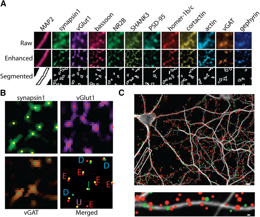
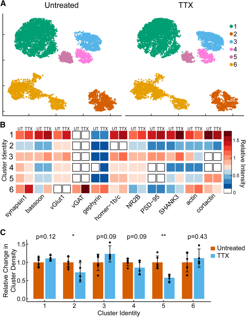
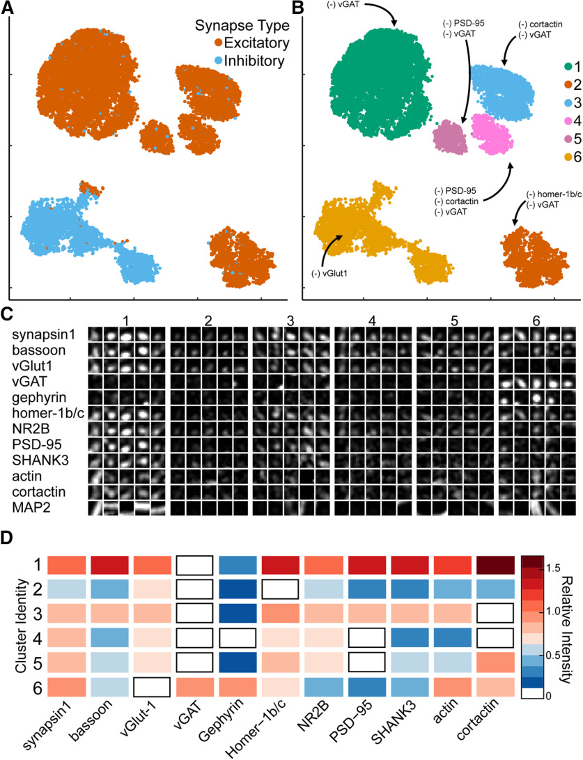
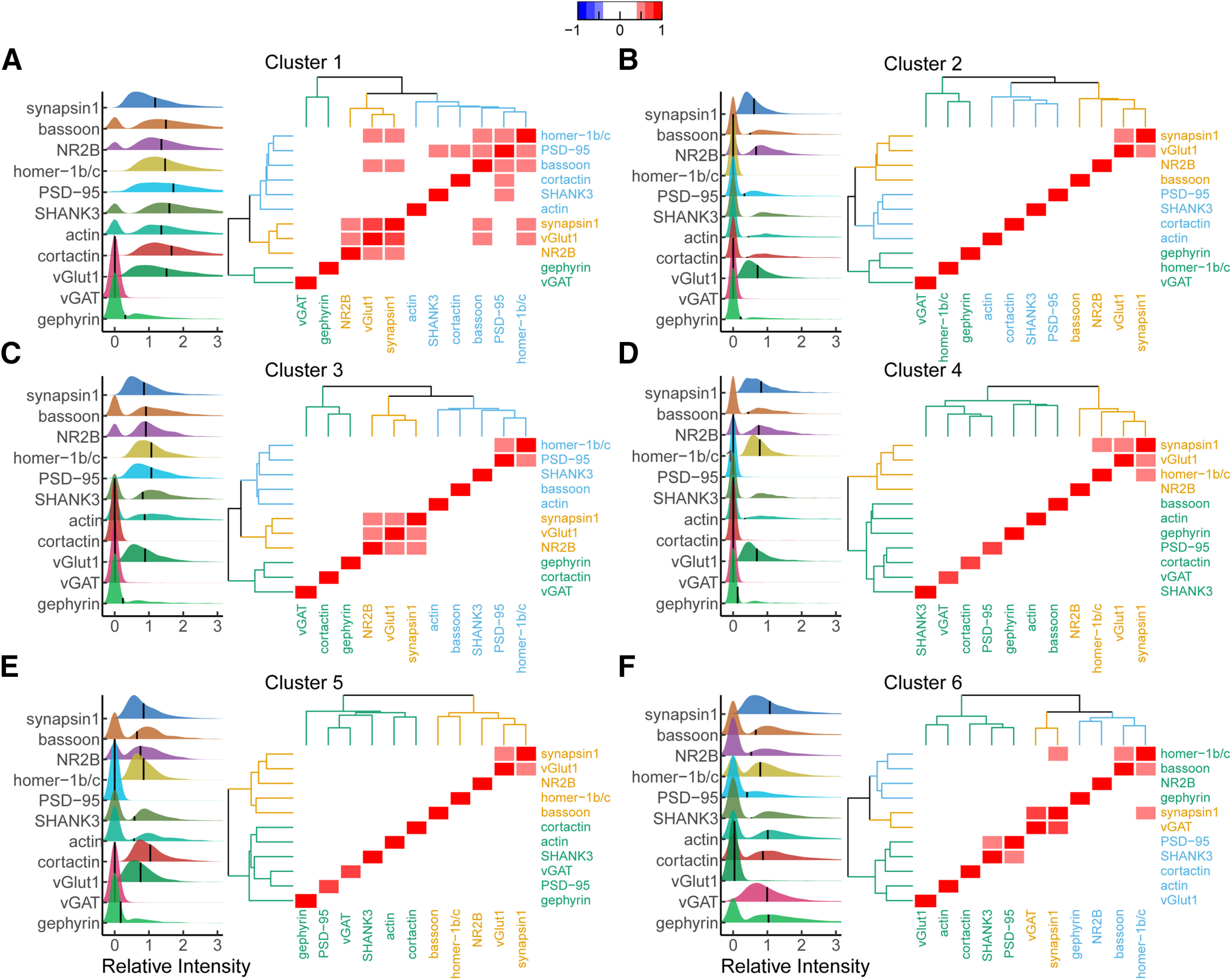
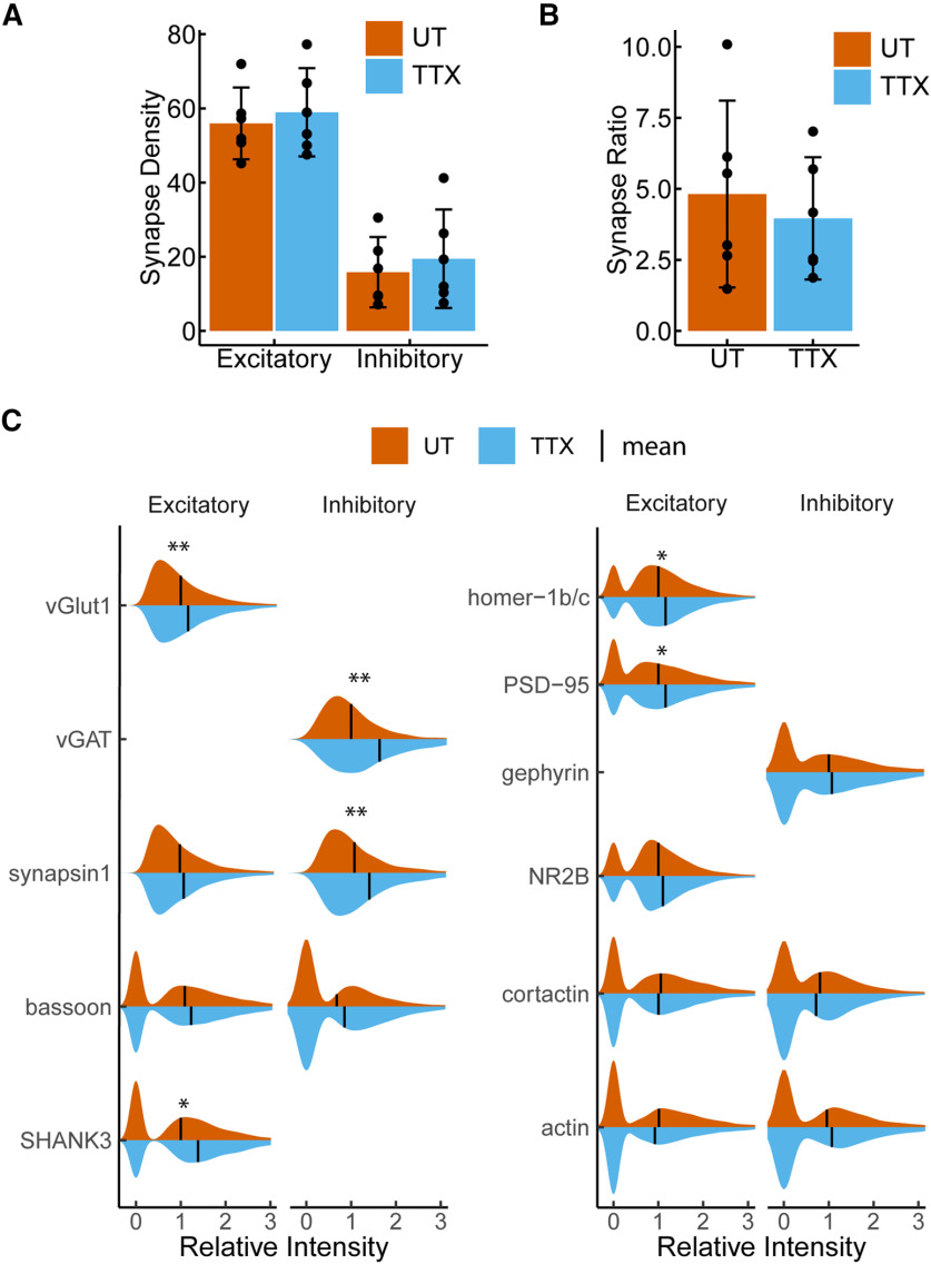
Neuronal synapses contain hundreds of different protein species important for regulating signal transmission. Characterizing differential expression profiles of proteins within synapses in distinct regions of the brain has revealed a high degree of synaptic diversity defined by unique molecular organization. Multiplexed imaging of rat primary hippocampal culture models at single synapse resolution offers new opportunities for exploring synaptic reorganization in response to chemical and genetic perturbations. Here, we combine 12-color multiplexed fluorescence imaging with quantitative image analysis and machine learning to identify novel synaptic subtypes within excitatory and inhibitory synapses based on the expression profiles of major synaptic components. We characterize differences in the correlated expression of proteins within these subtypes and we examine how the distribution of these synapses is modified following induction of synaptic plasticity. Under chronic suppression of neuronal activity, phenotypic characterization revealed coordinated increases in both excitatory and inhibitory protein levels without changes in the distribution of synaptic subtypes, suggesting concerted events targeting glutamatergic and GABAergic synapses. Our results offer molecular insight into the mechanisms of synaptic plasticity.
神经元突触包含数百种不同的蛋白质物种,这些蛋白质对于调节信号传递很重要。对大脑不同区域突触内蛋白质的差异表达谱进行特征描述,揭示了高度多样化的突触,其定义为独特的分子组织。在单个突触分辨率下对大鼠原代海马培养模型进行的 12 色多重荧光成像为探索化学和遗传干扰下的突触重组提供了新的机会。在这里,我们结合 12 色多重荧光成像与定量图像分析和机器学习,根据主要突触成分的表达谱,在兴奋性和抑制性突触内识别新型突触亚型。我们描述了这些亚型内蛋白质的相关表达的差异,并研究了在诱导突触可塑性后这些突触的分布如何被改变。在神经元活动的慢性抑制下,表型特征显示兴奋性和抑制性蛋白水平同时升高,而突触亚型的分布没有变化,这表明针对谷氨酸能和 GABA 能突触的协同事件。我们的结果为突触可塑性的机制提供了分子见解。