Sorbonne Université, Centre National de la Recherche Scientifique, Institut de Biologie Paris-Seine,, France.
Université de Limoges, XLIM, CNRS UMR 7252, Limoges, France.
J Biomed Opt. 2019 Oct;24(10):1-12. doi: 10.1117/1.JBO.24.10.106004.
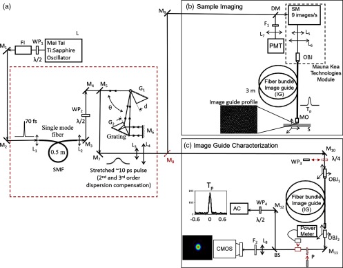
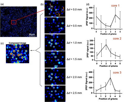
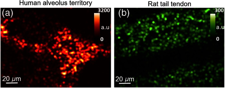
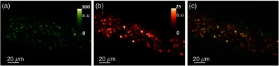
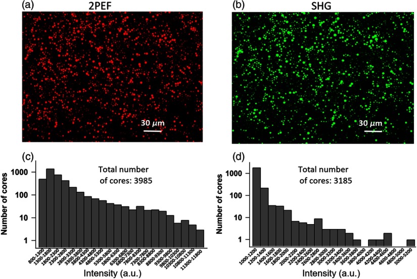
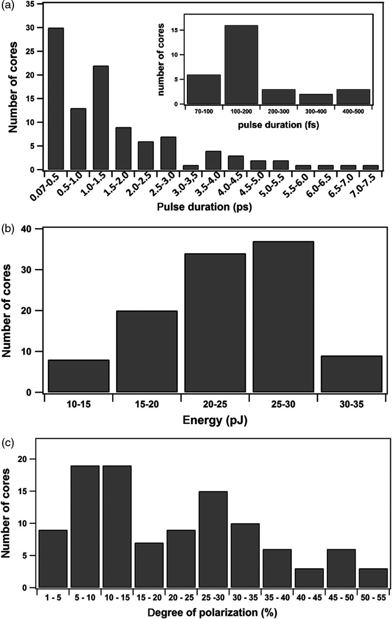
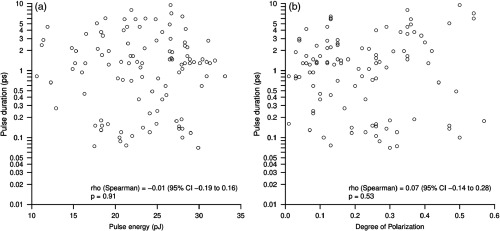
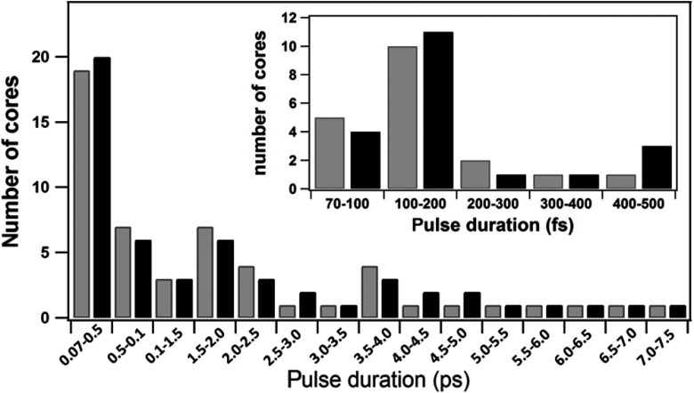
Multiphoton microscopy (MPM) has the capacity to record second-harmonic generation (SHG) and endogenous two-photon excitation fluorescence (2PEF) signals emitted from biological tissues. The development of fiber-based miniaturized endomicroscopes delivering pulses in the femtosecond range will allow the transfer of MPM to clinical endoscopy. We present real-time SHG and 2PEF ex vivo images using an endomicroscope, which totally complies with clinical endoscopy regulations. This system is based on the proximal scanning of a commercial multicore image guide (IG). For understanding the inhomogeneities of the recorded images, we quantitatively characterize the IG at the single-core level during nonlinear excitation. The obtained results suggest that these inhomogeneities originate from the variable core geometries that, therefore, exhibit variable nonlinear and dispersive properties. Finally, we propose a method based on modulation of dispersion precompensation to address the image inhomogeneity issue and, as a proof of concept, we demonstrate its capability to improve the nonlinear image quality.
多光子显微镜(MPM)能够记录从生物组织中发射的二次谐波产生(SHG)和内双光子激发荧光(2PEF)信号。基于光纤的小型化内窥镜在飞秒范围内传输脉冲,将 MPM 技术应用于临床内窥镜检查。我们使用内窥镜实时显示 SHG 和 2PEF 的离体图像,该内窥镜完全符合临床内窥镜检查规定。该系统基于对商业多芯图像引导器(IG)的近端扫描。为了理解记录图像的不均匀性,我们在非线性激发过程中对单芯水平的 IG 进行定量表征。结果表明,这些不均匀性源于可变的核心几何形状,因此表现出可变的非线性和色散特性。最后,我们提出了一种基于色散预补偿调制的方法来解决图像不均匀性问题,并作为概念验证,我们证明了它能够提高非线性图像质量的能力。