Maneas Efthymios, Aughwane Rosalind, Huynh Nam, Xia Wenfeng, Ansari Rehman, Kuniyil Ajith Singh Mithun, Hutchinson J Ciaran, Sebire Neil J, Arthurs Owen J, Deprest Jan, Ourselin Sebastien, Beard Paul C, Melbourne Andrew, Vercauteren Tom, David Anna L, Desjardins Adrien E
Wellcome/EPSRC Centre for Interventional and Surgical Sciences, University College London, London, UK.
Department of Medical Physics and Biomedical Engineering, University College London, London, UK.
J Biophotonics. 2020 Apr;13(4):e201900167. doi: 10.1002/jbio.201900167. Epub 2019 Nov 25.
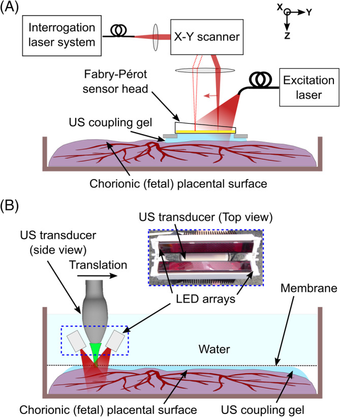
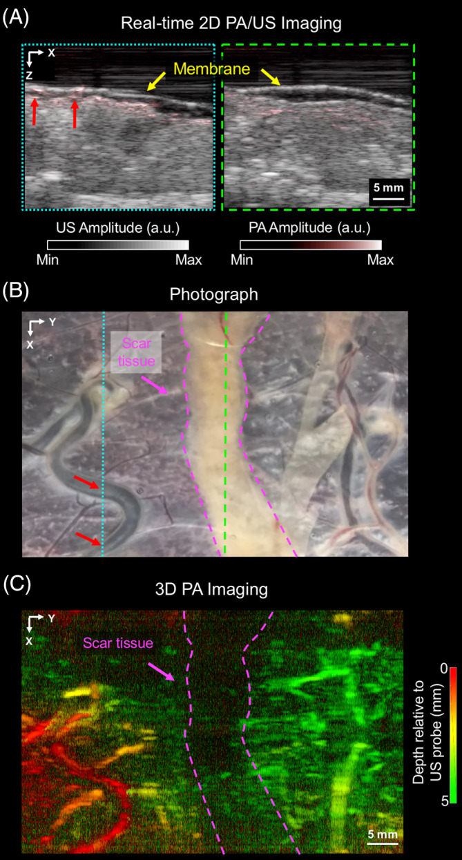
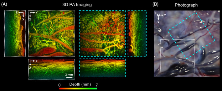
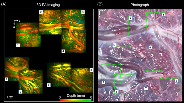
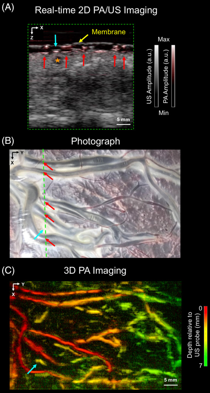
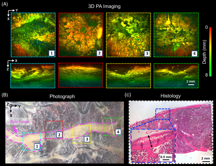
Minimally invasive fetal interventions require accurate imaging from inside the uterine cavity. Twin-to-twin transfusion syndrome (TTTS), a condition considered in this study, occurs from abnormal vascular anastomoses in the placenta that allow blood to flow unevenly between the fetuses. Currently, TTTS is treated fetoscopically by identifying the anastomosing vessels, and then performing laser photocoagulation. However, white light fetoscopy provides limited visibility of placental vasculature, which can lead to missed anastomoses or incomplete photocoagulation. Photoacoustic (PA) imaging is an alternative imaging method that provides contrast for hemoglobin, and in this study, two PA systems were used to visualize chorionic (fetal) superficial and subsurface vasculature in human placentas. The first system comprised an optical parametric oscillator for PA excitation and a 2D Fabry-Pérot cavity ultrasound sensor; the second, light emitting diode arrays and a 1D clinical linear-array ultrasound imaging probe. Volumetric photoacoustic images were acquired from ex vivo normal term and TTTS-treated placentas. It was shown that superficial and subsurface branching blood vessels could be visualized to depths of approximately 7 mm, and that ablated tissue yielded negative image contrast. This study demonstrated the strong potential of PA imaging to guide minimally invasive fetal therapies.
微创胎儿干预需要子宫腔内的精确成像。本研究中考虑的双胎输血综合征(TTTS)是由胎盘内异常的血管吻合引起的,这种吻合使得血液在胎儿之间不均匀流动。目前,TTTS通过胎儿镜检查来识别吻合血管,然后进行激光光凝治疗。然而,白光胎儿镜检查对胎盘血管系统的可视性有限,这可能导致吻合口遗漏或光凝不完全。光声(PA)成像是一种为血红蛋白提供对比度的替代成像方法,在本研究中,使用了两个PA系统来可视化人类胎盘的绒毛膜(胎儿)浅表和亚表面血管系统。第一个系统包括一个用于PA激发的光学参量振荡器和一个二维法布里-珀罗腔超声传感器;第二个系统包括发光二极管阵列和一个一维临床线性阵列超声成像探头。从体外足月胎盘和接受TTTS治疗的胎盘获取了体积光声图像。结果表明,浅表和亚表面分支血管可在约7毫米的深度内可视化,并且消融组织产生负图像对比度。本研究证明了PA成像在指导微创胎儿治疗方面的强大潜力。