• 文献检索
  • 文档翻译
  • 深度研究
  • 学术资讯
  • Suppr Zotero 插件Zotero 插件
  • 邀请有礼
  • 套餐&价格
  • 历史记录
应用&插件
Suppr Zotero 插件Zotero 插件浏览器插件Mac 客户端Windows 客户端微信小程序
定价
高级版会员购买积分包购买API积分包
服务
文献检索文档翻译深度研究API 文档MCP 服务
关于我们
关于 Suppr公司介绍联系我们用户协议隐私条款
关注我们

Suppr 超能文献

核心技术专利:CN118964589B侵权必究
粤ICP备2023148730 号-1Suppr @ 2026

文献检索

告别复杂PubMed语法,用中文像聊天一样搜索,搜遍4000万医学文献。AI智能推荐,让科研检索更轻松。

立即免费搜索

文件翻译

保留排版,准确专业,支持PDF/Word/PPT等文件格式,支持 12+语言互译。

免费翻译文档

深度研究

AI帮你快速写综述,25分钟生成高质量综述,智能提取关键信息,辅助科研写作。

立即免费体验

基于局部统计主动轮廓模型和结构先验的青光眼诊断中视网膜图像的视盘和杯区分割。

Optic Disc and Cup Segmentation in Retinal Images for Glaucoma Diagnosis by Locally Statistical Active Contour Model with Structure Prior.

机构信息

College of Computer Science, Shenyang Aerospace University, Shenyang, Liaoning, China.

School of Software, Jiangxi Normal University, Nanchang, Jiangxi, China.

出版信息

Comput Math Methods Med. 2019 Nov 20;2019:8973287. doi: 10.1155/2019/8973287. eCollection 2019.

DOI:10.1155/2019/8973287
PMID:31827591
原文链接:https://pmc.ncbi.nlm.nih.gov/articles/PMC6886352/
Abstract

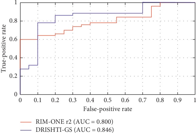
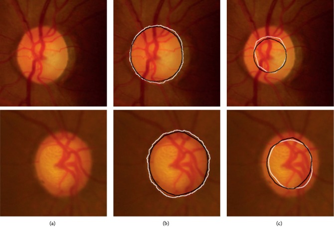
Accurate optic disc and optic cup segmentation plays an important role for diagnosing glaucoma. However, most existing segmentation approaches suffer from the following limitations. On the one hand, image devices or illumination variations always lead to intensity inhomogeneity in the fundus image. On the other hand, the spatial prior knowledge of optic disc and optic cup, e.g., the optic cup is always contained inside the optic disc region, is ignored. Therefore, the effectiveness of segmentation approaches is greatly reduced. Different from most previous approaches, we present a novel locally statistical active contour model with the structure prior (LSACM-SP) approach to jointly and robustly segment the optic disc and optic cup structures. First, some preprocessing techniques are used to automatically extract initial contour of object. Then, we introduce the locally statistical active contour model (LSACM) to optic disc and optic cup segmentation in the presence of intensity inhomogeneity. Finally, taking the specific morphology of optic disc and optic cup into consideration, a novel structure prior is proposed to guide the model to generate accurate segmentation results. Experimental results demonstrate the advantage and superiority of our approach on two publicly available databases, i.e., DRISHTI-GS and RIM-ONE r2, by comparing with some well-known algorithms.

摘要

准确的视盘和视杯分割对视神经疾病的诊断起着重要作用。然而,大多数现有的分割方法都存在以下局限性。一方面,图像设备或照明变化总会导致眼底图像的强度不均匀。另一方面,视盘和视杯的空间先验知识(例如,视杯总是包含在视盘区域内)被忽略了。因此,分割方法的有效性大大降低。与大多数先前的方法不同,我们提出了一种新的基于局部统计主动轮廓模型和结构先验的方法(LSACM-SP),用于联合且稳健地分割视盘和视杯结构。首先,采用一些预处理技术来自动提取目标的初始轮廓。然后,我们在存在强度不均匀的情况下引入局部统计主动轮廓模型(LSACM)进行视盘和视杯分割。最后,考虑到视盘和视杯的特定形态,提出了一种新的结构先验来指导模型生成准确的分割结果。实验结果表明,与一些知名算法相比,我们的方法在两个公开可用的数据库(即 DRISHTI-GS 和 RIM-ONE r2)上具有优势和优越性。

https://cdn.ncbi.nlm.nih.gov/pmc/blobs/0ea9/6886352/f4d86529b370/CMMM2019-8973287.011.jpg
https://cdn.ncbi.nlm.nih.gov/pmc/blobs/0ea9/6886352/dde23618ea3c/CMMM2019-8973287.001.jpg
https://cdn.ncbi.nlm.nih.gov/pmc/blobs/0ea9/6886352/30207584db25/CMMM2019-8973287.002.jpg
https://cdn.ncbi.nlm.nih.gov/pmc/blobs/0ea9/6886352/03a66e500a0d/CMMM2019-8973287.003.jpg
https://cdn.ncbi.nlm.nih.gov/pmc/blobs/0ea9/6886352/6b2177996e5d/CMMM2019-8973287.004.jpg
https://cdn.ncbi.nlm.nih.gov/pmc/blobs/0ea9/6886352/e353b5259dde/CMMM2019-8973287.005.jpg
https://cdn.ncbi.nlm.nih.gov/pmc/blobs/0ea9/6886352/7034f78e4ce0/CMMM2019-8973287.006.jpg
https://cdn.ncbi.nlm.nih.gov/pmc/blobs/0ea9/6886352/26e3ade872bd/CMMM2019-8973287.007.jpg
https://cdn.ncbi.nlm.nih.gov/pmc/blobs/0ea9/6886352/a8bfa53a7f9b/CMMM2019-8973287.008.jpg
https://cdn.ncbi.nlm.nih.gov/pmc/blobs/0ea9/6886352/d43d13989903/CMMM2019-8973287.009.jpg
https://cdn.ncbi.nlm.nih.gov/pmc/blobs/0ea9/6886352/1cffa7c7802f/CMMM2019-8973287.010.jpg
https://cdn.ncbi.nlm.nih.gov/pmc/blobs/0ea9/6886352/f4d86529b370/CMMM2019-8973287.011.jpg

相似文献

1
Optic Disc and Cup Segmentation in Retinal Images for Glaucoma Diagnosis by Locally Statistical Active Contour Model with Structure Prior.基于局部统计主动轮廓模型和结构先验的青光眼诊断中视网膜图像的视盘和杯区分割。
Comput Math Methods Med. 2019 Nov 20;2019:8973287. doi: 10.1155/2019/8973287. eCollection 2019.
2
Fully automated method for glaucoma screening using robust optic nerve head detection and unsupervised segmentation based cup-to-disc ratio computation in retinal fundus images.基于视网膜眼底图像中稳健的视神经头检测和无监督视杯/视盘比计算的青光眼筛查全自动方法。
Comput Med Imaging Graph. 2019 Oct;77:101643. doi: 10.1016/j.compmedimag.2019.101643. Epub 2019 Aug 14.
3
Fully Convolutional Networks for Monocular Retinal Depth Estimation and Optic Disc-Cup Segmentation.基于全卷积网络的单目视网膜深度估计和视盘-杯分割
IEEE J Biomed Health Inform. 2019 Jul;23(4):1417-1426. doi: 10.1109/JBHI.2019.2899403. Epub 2019 Feb 14.
4
Joint optic disc and cup segmentation based on densely connected depthwise separable convolution deep network.基于深度可分离卷积深度网络的联合视盘和杯分割。
BMC Med Imaging. 2021 Jan 28;21(1):14. doi: 10.1186/s12880-020-00528-6.
5
Patch-Based Output Space Adversarial Learning for Joint Optic Disc and Cup Segmentation.基于补丁的输出空间对抗学习在视盘和杯联合分割中的应用。
IEEE Trans Med Imaging. 2019 Nov;38(11):2485-2495. doi: 10.1109/TMI.2019.2899910. Epub 2019 Feb 18.
6
Optic Disc and Cup Image Segmentation Utilizing Contour-Based Transformation and Sequence Labeling Networks.利用基于轮廓的变换和序列标注网络进行视盘和杯图像分割。
J Med Syst. 2020 Mar 20;44(5):96. doi: 10.1007/s10916-020-01561-2.
7
Accurate, fast, data efficient and interpretable glaucoma diagnosis with automated spatial analysis of the whole cup to disc profile.利用全自动的视杯-视盘整体形态分析实现准确、快速、高效的数据利用和可解释的青光眼诊断。
PLoS One. 2019 Jan 10;14(1):e0209409. doi: 10.1371/journal.pone.0209409. eCollection 2019.
8
Automatic optic disc segmentation with peripapillary atrophy elimination.具有消除视乳头周围萎缩功能的自动视盘分割
Annu Int Conf IEEE Eng Med Biol Soc. 2011;2011:6224-7. doi: 10.1109/IEMBS.2011.6091537.
9
Rim-to-Disc Ratio Outperforms Cup-to-Disc Ratio for Glaucoma Prescreening.边缘到盘比优于杯盘比用于青光眼初筛。
Sci Rep. 2019 May 8;9(1):7099. doi: 10.1038/s41598-019-43385-2.
10
A novel lightweight deep learning approach for simultaneous optic cup and optic disc segmentation in glaucoma detection.一种用于青光眼检测的新型轻量级深度学习方法,用于同时分割视杯和视盘。
Math Biosci Eng. 2024 Mar 4;21(4):5092-5117. doi: 10.3934/mbe.2024225.

引用本文的文献

1
A residual connection enabled deep neural network model for optic disk and optic cup segmentation for glaucoma diagnosis.基于残差连接的深度神经网络模型用于青光眼诊断的视盘和视杯分割。
Sci Prog. 2023 Jul-Sep;106(3):368504231201329. doi: 10.1177/00368504231201329.
2
Clinical Applications of Artificial Intelligence in Glaucoma.人工智能在青光眼领域的临床应用
J Ophthalmic Vis Res. 2023 Feb 21;18(1):97-112. doi: 10.18502/jovr.v18i1.12730. eCollection 2023 Jan-Mar.
3
Detection of Optic Disc Localization from Retinal Fundus Image Using Optimized Color Space.

本文引用的文献

1
Adaptive weighted locality-constrained sparse coding for glaucoma diagnosis.基于自适应加权局部约束稀疏编码的青光眼诊断。
Med Biol Eng Comput. 2019 Sep;57(9):2055-2067. doi: 10.1007/s11517-019-02011-z. Epub 2019 Jul 27.
2
Robust optic disc and cup segmentation with deep learning for glaucoma detection.利用深度学习进行青光眼检测的稳健视盘和杯分割。
Comput Med Imaging Graph. 2019 Jun;74:61-71. doi: 10.1016/j.compmedimag.2019.02.005. Epub 2019 Apr 5.
3
Automatic Optic Disc Segmentation Based on Modified Local Image Fitting Model with Shape Prior Information.
利用优化颜色空间检测视网膜眼底图像中的视盘定位。
J Digit Imaging. 2022 Apr;35(2):302-319. doi: 10.1007/s10278-021-00566-8. Epub 2022 Jan 11.
4
Segmentation of Intensity-Corrupted Medical Images Using Adaptive Weight-Based Hybrid Active Contours.基于自适应权重的混合主动轮廓线对强度污染医学图像的分割。
Comput Math Methods Med. 2020 Nov 4;2020:6317415. doi: 10.1155/2020/6317415. eCollection 2020.
基于改进的局部图像拟合模型和形状先验信息的自动视盘分割。
J Healthc Eng. 2019 Mar 14;2019:2745183. doi: 10.1155/2019/2745183. eCollection 2019.
4
Joint Optic Disc and Cup Segmentation Based on Multi-Label Deep Network and Polar Transformation.基于多标签深度网络和极坐标变换的联合视盘和杯分割。
IEEE Trans Med Imaging. 2018 Jul;37(7):1597-1605. doi: 10.1109/TMI.2018.2791488.
5
A Novel Adaptive Deformable Model for Automated Optic Disc and Cup Segmentation to Aid Glaucoma Diagnosis.一种用于自动视盘和杯分割以辅助青光眼诊断的新型自适应可变形模型。
J Med Syst. 2017 Dec 7;42(1):20. doi: 10.1007/s10916-017-0859-4.
6
Detection of Dendritic Spines Using Wavelet Packet Entropy and Fuzzy Support Vector Machine.基于小波包熵和模糊支持向量机的树突棘检测。
CNS Neurol Disord Drug Targets. 2017;16(2):116-121. doi: 10.2174/1871527315666161111123638.
7
A Level Set Approach to Image Segmentation With Intensity Inhomogeneity.基于水平集的强度不均匀图像分割方法。
IEEE Trans Cybern. 2016 Feb;46(2):546-57. doi: 10.1109/TCYB.2015.2409119. Epub 2015 Mar 12.
8
Sparse dissimilarity-constrained coding for glaucoma screening.用于青光眼筛查的稀疏差异约束编码
IEEE Trans Biomed Eng. 2015 May;62(5):1395-403. doi: 10.1109/TBME.2015.2389234. Epub 2015 Jan 9.
9
Trainable COSFIRE filters for vessel delineation with application to retinal images.可训练的 COSFIRE 滤波器在视网膜图像中的血管分割应用
Med Image Anal. 2015 Jan;19(1):46-57. doi: 10.1016/j.media.2014.08.002. Epub 2014 Sep 3.
10
Superpixel classification based optic disc and optic cup segmentation for glaucoma screening.基于超像素分类的青光眼筛查的视盘和视杯分割。
IEEE Trans Med Imaging. 2013 Jun;32(6):1019-32. doi: 10.1109/TMI.2013.2247770. Epub 2013 Feb 18.