Matsumoto Celso Soiti, Shibuya Masayuki, Makita Jun, Shoji Takuhei, Ohno Hisato, Shinoda Kei, Matsumoto Harue
Department of Ophthalmology, Teikyo University School of Medicine, Kaga 2-11-1, Itabashi-ku, Tokyo 173-8605, Japan.
Department of Ophthalmology, Saitama Medical University School of Medicine, Morohongo 38, Moroyama-machi, Saitama 350-0495, Japan.
J Ophthalmol. 2019 Nov 25;2019:5013463. doi: 10.1155/2019/5013463. eCollection 2019.
To determine the feasibility of performing intraocular surgeries in a heads-up position with low illuminance conditions by observing a display of the surgical field created by a three-dimensional imaging (3D) system.
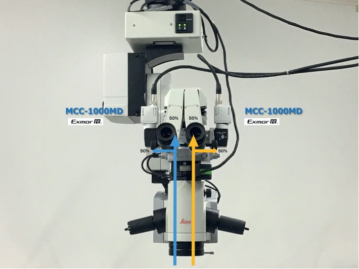
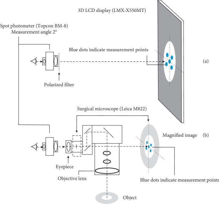
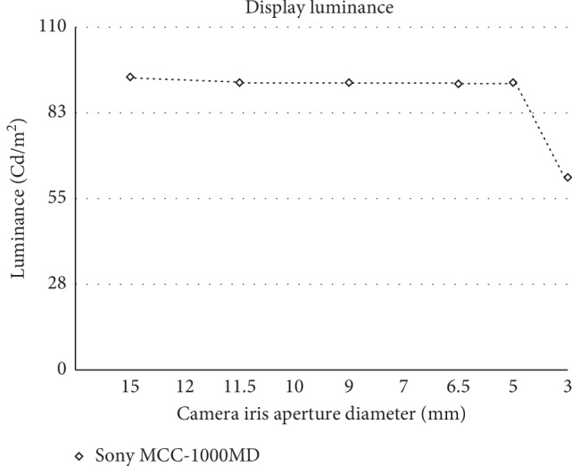
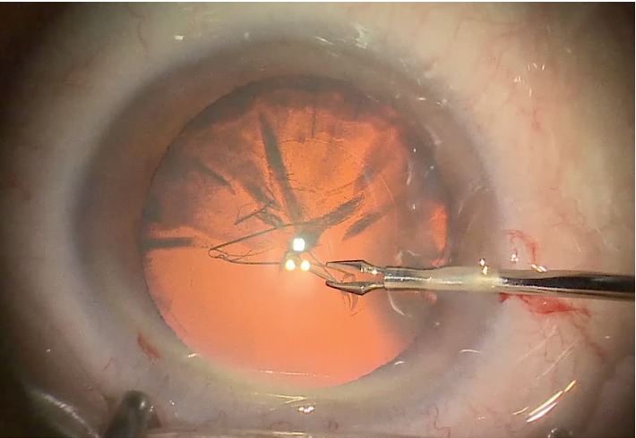
Seventy-four eyes of 56 patients underwent cataract surgery (72 eyes) with the heads-up 3D surgery system; 60 eyes with cataract surgery alone, 7 eyes with combined cataract and glaucoma microdevice implant surgery, 5 eyes with combined cataract and vitrectomy surgery, and two eyes with vitrectomy surgery alone were studied. The illuminance from the surgical microscope was set to be dimmer (Leica M822F40 main light 2%; otto-flex 6%) than the usual setting to minimize the discomfort and glare for the patient. The surgeries were performed under topical anesthesia. The luminance of the images observed through the eyepieces of the operating microscope and the image of a 3D system created by a high-sensitivity sensor Exmor R 3CMOS HD camera (Sony MCC-1000MD) were measured.
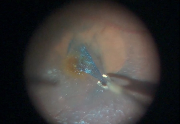
All surgeries were completed without any complications under the low illumination conditions. The surgical field on the display monitor was created by a 3D system using a high-sensitivity sensor camera and was observed in a heads-up position. The patients did not report any intolerable discomfort or glare during the surgery. Cataract surgeries were performed with a good view of the surgical field under the extremely low illumination from the surgical microscope. The high-sensitivity sensors and electronic amplifications of the image signals made the surgical field brighter and allowed the surgeon to perform the surgery confidently and safely.
Heads-up, 3D-assisted intraocular surgeries can be performed safely and efficiently with low illuminance of the surgical field. This trial is registered with UMIN000037838.
通过观察三维成像(3D)系统创建的手术视野显示屏,确定在低光照条件下抬头位进行眼内手术的可行性。
56例患者的74只眼接受了使用抬头式3D手术系统的白内障手术(72只眼);研究了60只仅行白内障手术的眼、7只白内障合并青光眼微装置植入手术的眼、5只白内障合并玻璃体切割手术的眼以及2只仅行玻璃体切割手术的眼。手术显微镜的光照度设置得比通常情况更暗(徕卡M822F40主灯2%;奥托 - 弗莱克斯6%),以尽量减少患者的不适和眩光。手术在表面麻醉下进行。测量了通过手术显微镜目镜观察到的图像亮度以及由高灵敏度传感器Exmor R 3CMOS HD相机(索尼MCC - 1000MD)创建的3D系统图像的亮度。
所有手术均在低光照条件下顺利完成,无任何并发症。显示屏上的手术视野由使用高灵敏度传感器相机的3D系统创建,并在抬头位观察。患者在手术过程中未报告任何无法忍受的不适或眩光。在手术显微镜极低光照条件下进行白内障手术时,手术视野清晰。图像信号的高灵敏度传感器和电子放大使手术视野更亮,使外科医生能够自信且安全地进行手术。
在手术视野低光照的情况下,抬头位三维辅助眼内手术能够安全、高效地进行。本试验已在UMIN000037838注册。